Exploring the cultivated silk moth Bombyx mori.
Part II: Further microscopical studies of aspects of the life cycle with notes on Leeuwenhoek's observations.
by David Walker, UK
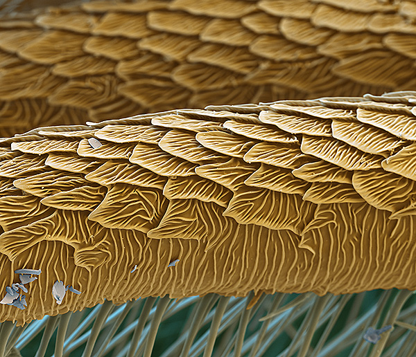
SEM imagery courtesy of Oliver Meckes, 'Eyes of Science'.
In Explorer browser, setting 60% in 'Print Preview' prints out on 9 sheets of A4 paper.
Part 1 (June 2012 Micscape) - adult wing, scales and scale fine structure.
Part 3 (October 2012 Micscape) - compound eyes of adult, facet count.
In part I (June 2012 Micscape issue), as a hobbyist, I shared a microscopy study of the wings and scales of the adult cultivated silk moth Bombyx mori; the modern microscope with its fully corrected optics and wealth of contrast enhancement techniques revealed little that Leeuwenhoek's detailed studies with single lens microscopes did not reveal. In this part II, other aspects of the life cycle of Bombyx mori are explored. The silk moth's long history in sericulture and being one of the earliest insects to be studied in detail gives it a particular appeal for the enthusiast to study under the modern microscope. By manipulating and studying the same subjects, an additional insight and appreciation can be gained of the skills of the early workers such as Leeuwenhoek, Malpighi and Swammerdam (1, 1a).
Studies by Leeuwenhoek
Antoni van Leeuwenhoek's studies of the silk moth and its life cycle as catalogued by Cole (2) were mainly discussed in two letters; English translations of both are available in Hoole's 'Select Works' (3) and the definitive 'Collected Letters' (4).
Letter dated July 11th 1687 to the Royal Society - this was not published in the Society's Philosophical Transactions. It describes his detailed studies of the eggs and of the larva within the egg at various stages of development. He took great care in keeping the eggs warm in colder weather and with admirable support from his wife! Leeuwenhoek notes (3):
"Some of these eggs, which were six weeks old, I put into a flat screwed box, which in the day time I carried in my pocket, and at night placed beside me in bed, that they may be continually kept warm : and in another box of the same kind, I put some more eggs, three weeks old, and these my wife (who was always very warmly clad) constantly carried in her bosom. This we did, to try the experiment, whether it were possible to promote the growth of Silk-worms in the autumn."
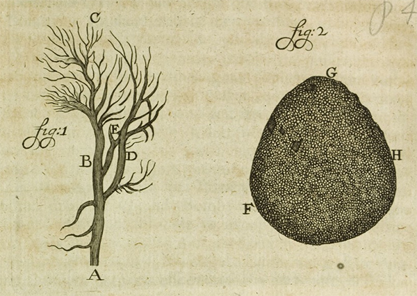
The structure of the trachea in the larva was also described and illustrated. The 'Collected Letters' notes that the original two figures sent to the Royal Society were lost but they are shown in contemporary published editions of the Letters. The plate below is from 'Opera Omnia', a Latin edition.
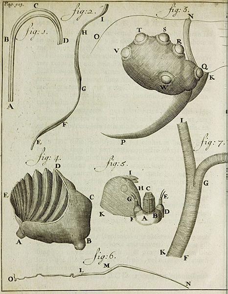
Plate to accompany Leeuwenhoek's letter dated July 11th 1687. Fig. 1 shows the trachea (Leeuwenhoek's 'vessels') on one side of the head of an unborn silkworm. Fig. 2 is an egg, 'GH' is where the silkworm has bitten through to emerge.
Plate from the published letters in 'Opera Omnia', vol. 1, 1722. See original plate on 'ECHO' website (European Cultural Heritage Online).
ECHO credit and 'source information' for this plate and plates below: Digitised by and images © Max Planck Institute for the History of Science. The ECHO venture has an 'Open Access Policy' where public display of works including derivative works is permitted with proper attribution.
Letter dated April 20th 1702 to Karl, Landgrave of Hessen-Kassel. This described studies of silkworm structure, the silk thread and the organs producing it. The letter also included detailed studies of the adult wings and wing scale structure including accurate measurements of the fine structure of the wing scales which were presented in the earlier Micscape article.
|
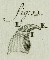
Right: Plate to accompany Leeuwenhoek's letter dated 20th April 1702. Plate from the published letters in 'Opera Omnia', vol. 4, 1719. See original plate. Image credit - see earlier plate. Fig. 1 - twin filaments of the silk thread which sometimes partially separate. Fig. 2 - the flattened profile of the thread caused by the twinning. Fig. 3 - side of the silkworm head showing the six simple eyes, now called lateral ocelli or stemmata (7). Fig. 4 - one of the two mandibles. Fig. 5 - the labium underside; DE and FG are labial palps, C the spinneret, HIK is the right mandible (as described in the modern caption to the 'Collected Letters' plate.) These features can be compared to the SEM of the silkworm shown below. Fig. 6 - a silk gland. Fig. 7 - part of the trachea. |
|
Studying the life cycle of Bombyx mori
The cultivated silk moth is a popular subject in education and a useful resource is the plastic embedded complete lifecycle available on eBay and various suppliers. They are not suited for detailed microscopic studies but work well enough for visual or low power study of major features, although the embedding alters the true surface texture, notably the wings of the adult which become transparent.
A number of suppliers offer rearing kits from eggs but does need a commitment of time and resources to feed and tend until adult. I have incubated viable cocoons to adult as the moths do not feed and only live for a short time after mating (see part I).
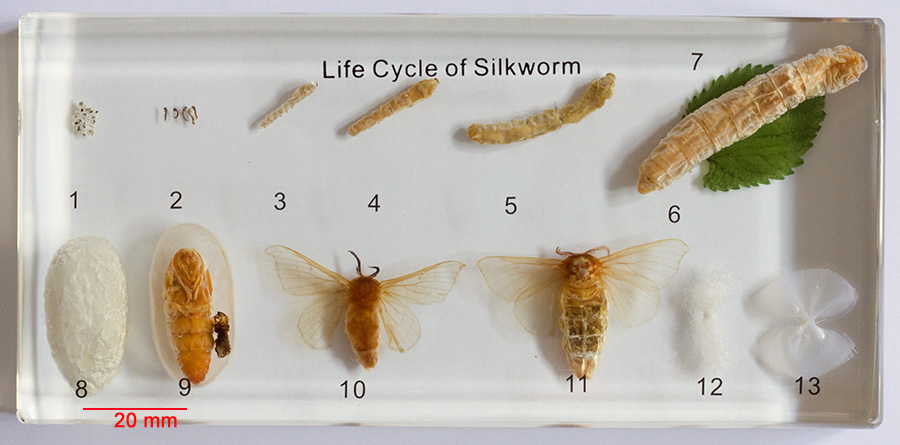
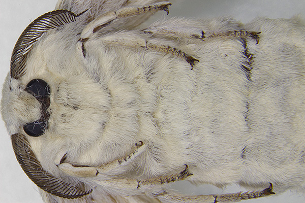
Note the more robust adult female on right, although the reported differences between male and female antennae were not that distinct in this pair. Only females hatched from my own viable cocoons, where the gland to release the pheromones was very distinct (see Part I). Suspected males were sorted from shrivelled dead specimens for antenna study below, but when shrivelled, differences between sexes seemed less distinct.
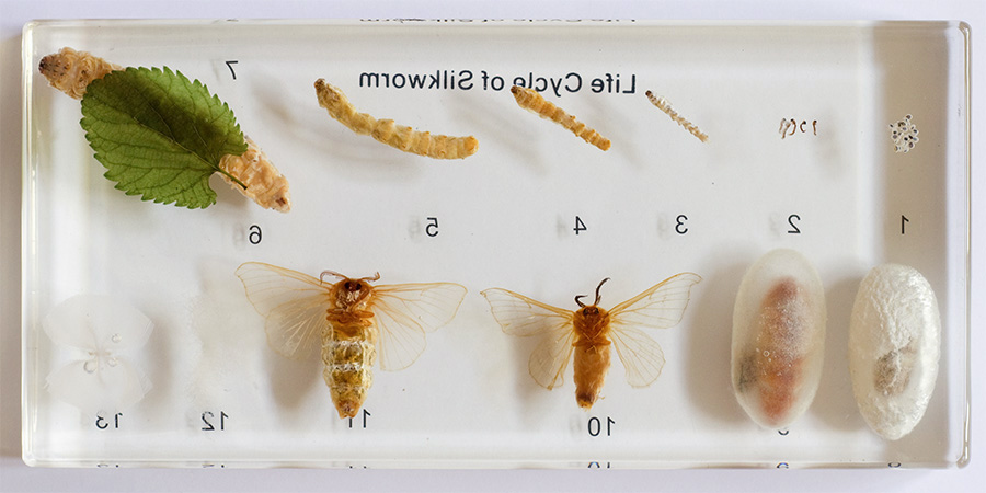
Images above, the widely available plastic embedded life cycle of Bombyx mori with supplied laminated index. An advantage of this presentation is the ability to study each stage in the life cycle from above and below.
Head and legs - studied using reared adults
|
A female silk moth Bombyx mori reared from a viable cocoon is shown right. The double claws on the legs are seen which Leeuwenhoek described and illustrated. See inset below. Stereo microscope, optical mag 5X.
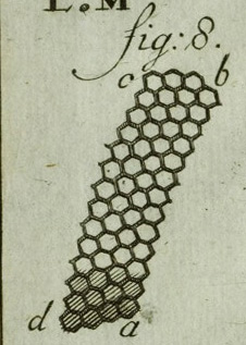
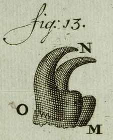
Above: Fig. 8, facets of adult eye and fig. 13, detail of silk moth leg claw. From plate to accompany Leeuwenhoek's letter dated 20th April 1702. Plate from the published letters in 'Opera Omnia', vol. 4, 1719. See original plate. Image credit - see earlier plate. |
Horizontal field width (HFW) = 15.8 mm |
||
|
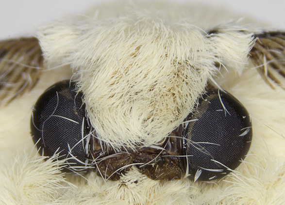
Head detail of a female showing the typical compound eyes and the extensive covering of scales / scale like hairs. The eyes of the adult silk moth were the subject of one of Leeuwenhoek's famous micrometric studies and described in his letter dated 20th April 1702. He counted the facets across a circumference and in his letter shows his arithmetical calculations to derive an estimate for the total number of facets. Stereo microscope, optical mag 10X. |
Horizontal field width (HFW) = ca. 4 mm |
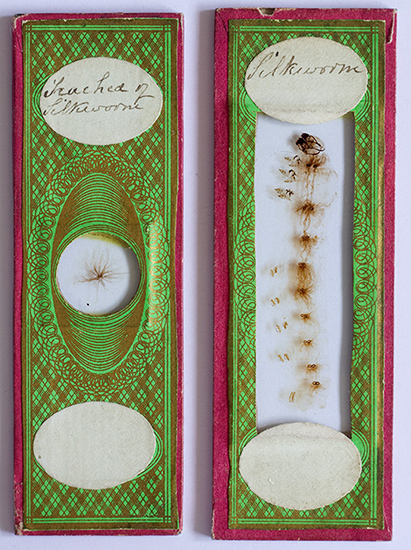 Larva and trachea - studied using two Victorian papered slides
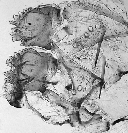
The life cycle of the silk moth often formed the subject of antique slides and two attractive papered slides possessed are shown right; both are unnamed and undated but are typical papered slides of the last half of the 19th century. Both mounts look as clear as the day they were made. (Comments on the likely slide maker would be of interest.)
One is a splendid mount of the whole silkworm and another of the dissected and laid out 'trachea' (i.e. both of the larval stage). Modern slides of the life cycle can often be obtained from major slide makers e.g. Lieder.
Prepared insect slides such as these are useful for studies of finer structures but really need to be complemented by studies of the undistorted unmounted subject which correctly presents the three dimensionality and spatial arrangement of features. Compare for example the false layout of the head features in the prepared slide with that of the SEM image below it.
Imaging the whole silkworm slide with a compound microscope would involve a number of image stitches but an easier way, very suited to large slide mounts, is to use a 35 mm slide film scanner. Although the silkworm's size still required two stitches as it's wider than the 35 mm film format. The scanning techniques used have been discussed in earlier articles.
An image of the whole silkworm scanned with a Minolta Dimage Elite 5400 dpi scanner is shown below, considerably resized from the 20 Mpixel original. Further images of the larval details under the compound microscope are then shown.
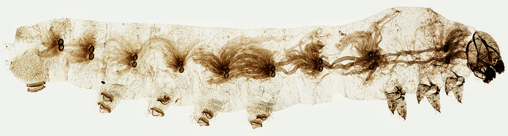
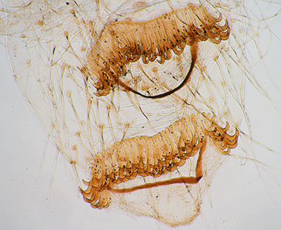
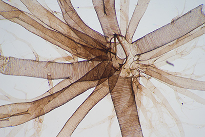
Silkworm, papered microscope slide, scanned directly with a 35 mm slide film scanner. The silkworm (length 42 mm) shows features typical of that for the Lepidopteran larva (Imms in his classic 'A General Textbook of Entomology' uses Bombyx mori as the illustrated example, 5). There are three pairs of thoracic legs, five pairs of abdominal legs 'pro-legs' and tracheal structures leading to nine pairs of spiracles are clearly seen.
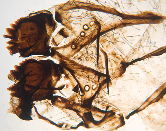
Close up of the larval mouthparts, Zeiss 2.5X objective. Left, visible brightfield; right; near infrared brightfield using a 760 nm filter on the field iris of a Zeiss Photomicroscope. The mandibles and lateral ocelli can be clearly seen. HFW=3.5 mm for lefthand image.
Near IR can penetrate denser areas of insect exoskeletons and can be useful for showing detail. Out of camera images are tonally flat so tonal balance is required. The camera used was a home modified old Sony S75 consumer digicam described in this Micscape article. The camera is able to auto white balance avoiding the red tint of near IR imagery.
|
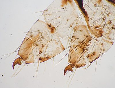
One of the three pairs of thoracic legs of the larva. This and following three images using a Zeiss 2.5X objective. HFW=2.4 mm. Right above: Fig. 12, detail of silkworm thoracic leg claw. From plate to accompany Leeuwenhoek's letter dated 20th April 1702. Plate from the published letters in 'Opera Omnia', vol. 4, 1719. See original plate. Image credit - see earlier plate. |
One of the five pairs of fleshy abdominal legs or pro-legs, lost in the adult. HFW=2.2 mm. |
|
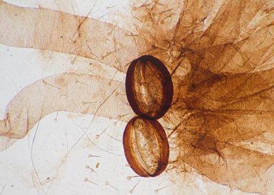
A pair of spiracles and part of the complex tracheal network. HFW=2 mm. |
A neat arrangement of part of the trachea on the second papered slide. The spiral thickening of the tubes can be seen. HFW=3 mm. |
|
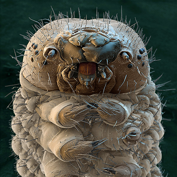
False colour SEM image of the head of a silkworm larva which beautifully shows the mandibles, lateral ocelli, spinneret and the thoracic legs. Image courtesy and copyright of Oliver Meckes of Eye of Science. Please contact them for image use. The exact function of the lateral ocelli (stemmata) in insect larvae that possess them is still debated and studied, see for example ref. 8. Leeuwenhoek's ability to accurately describe the complex features of the head (compare SEM with plate shown earlier) is an example of his micro-dissection skills. His silkworm studies included a number of the unborn larvae by opening an egg at various stages of development. Cole in his definitive review of Leeuwenhoek's zoological studies has a section on his micro-dissection work and eloquently remarks (2a): 'Leeuwenhoek's manipulative skills must have been astonishing, and he may certainly be acclaimed as the pioneer of micro-dissection. He has not told us how this work was carried out, and some of it is sufficiently difficult to tax a modern observer who has at hand all the refinements of modern binocular microscopes'.
|
|
Silk threads
Leeuwenhoek's studies of the silk thread were described and illustrated in his letter dated July 11th 1687 (see plate above). He accurately observed that each thread is comprised of two finer filaments glued together and which gives the thread a flattened profile of up to four times wider than broad. He correctly deduced that the lustre of silk cloth was related to the thread shape. Later studies have found that the threads have a roughly triangular cross section with rounded corners which are clearly
seen
under the SEM (see image by 'Eyes of Science' below). The prism shaped fibres contribute to the fibre's lustre by internal reflection of incident light.
|
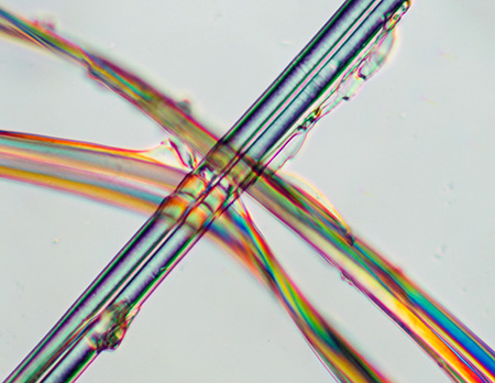
Untreated silk fibres teased from a cocoon. Zeiss 16X objective, DIC. Thread width 31 µm. Shows the twin filaments of each thread and the sericin protein coating of the central fibroin protein filaments. |
|
|
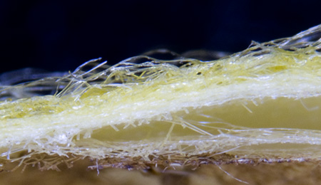
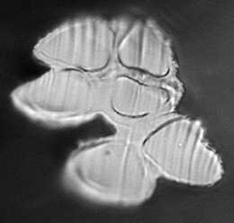
Edge on section of a cocoon. Optical mag 10X. Cocoon colour can vary between races. The cocoon right was a quite marked yellow, that below a much paler yellow. |
|
|
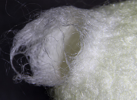
Emergence hole for adult in silk cocoon. Optical mag 5X. In silk production most pupae are not permitted to hatch as the exit hole of the adult moth damages the silk threads, shown right. The cocoons are typically heated to kill the pupae before emergence. The adult silk moth does not bite a hole to emerge but releases a liquid from its mouth that solubilises the cocoon. |
|
It's an interesting challenge to try and show the triangular cross section of the filaments under the optical microscope. One approach would be to embed a bundle of threads and cut cross sections with a microtome, but instead I tried cutting a thin strip of the stiff cocoon with a razor blade, as this should present a number of threads in rough cross section.
|
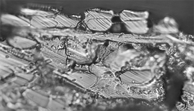
A cross section of the cocoon itself does not clearly show individual fibre structures. Epi 40/0.85 'pseudo DIC'. |
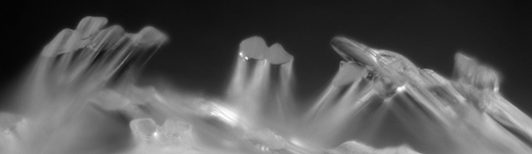
Scanning the loose fibres at cocoon edge does show an occasional clean section with the triangular shape, i.e. the central fibre. The twin nature of each thread is also shown. Epi with Zeiss 16/0.35 objective, crossed polars to reduced flare (as a none epi objective.) |
|
|
Image lower left. Three silk fibres showing their twinning and internal triangular structure with outer sheath. As with the top left-hand image, a Zeiss 40/0.85 epi DIC objective was used giving a 'pseudo DIC'. The reflectivity of the fibre ends was low with flare from surrounding fibres. Heavy post tonal balance adjustment was required to give a tolerable image. Width of central double filament = one silk thread ca. 28 µm. |
|
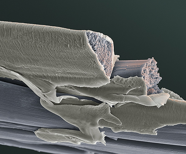
A false colour SEM of silk fibres in cross section more clearly shows the triangular structure and also the fibroin core and outer sheath of sericin, the latter glues the two filaments together. Image courtesy and copyright of Oliver Meckes of Eye of Science. Please contact them for image use. The sericin is usually removed by one of a number of hot water based treatments known as 'degumming' before further processing the silk. Potential uses of the waste sericin have been studied and developed and include its use in cosmetics and medical applications (6,7).
|
|
Antenna
|
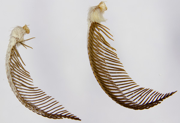
I haven't found references to date of Leeuwenhoek reporting on a study of the antennae of Bombyx mori. Antenna from (suspected) male and known female B. mori are shown right (stereo 10X optical mag.). The males are described as having denser sensory hairs but didn't find the difference that clear cut in the suspected males and known females I have had access to date, nor in the plastic embedded samples shown above. Antenna 5 mm high, width at max. ca. 1.2 mm. |
|
|
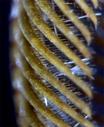
Close up of an antenna showing the finer hairs leading from each side 'rib'. Each 'rib' ca. 68 µm wide at max. (Stereo 30X optical mag.). To attract a male, the female releases pheromones from a gland on her abdomen. The predominant molecule is bombykol, structure below. It was the first pheromone to be isolated and chemical structure elucidated by Adolf Butenandt whose earlier work on human sex hormones was awarded the Nobel Prize in 1939 (but forbidden to accept it by the German government).
Bombykol
|
|
|
Detail of an antennae to show the surface structure and sensory hairs. Image courtesy and copyright of Oliver Meckes of Eye of Science. Please contact them for image use. My own attempts to show the surface structure of a 'rib' well on a compound microscope have been unsuccessful to date; axial epi with none epi objectives gave a lot of glare; hints of surface structure is just visible in the stereo image above. Intensive work by scientists has been carried out on how the male antennae on Bombyx and other insects detects these pheromones at such low concentrations, see for example ref. 9. Understanding how nature has designed very sensitive chemical receptors has inspired improved designs of manmade devices. See for example, 'Nanotool mimics moth antenna' article on Futurity website, with superb graphics showing how the B. mori antenna's oily coating to aid capture of the bombykol molecule was used to improve nanopore design. |
|
Comments to the author David Walker are welcomed.
Acknowledgements
I would like to thank the following people, but any errors in the above article are solely mine.
- William Hyett of www.silkfarm.co.uk for supplying the viable cocoons for hatching and for providing valuable advice on incubating them.
- The Science Museum Library (London) staff for the copy of Leeuwenhoek's Letters in references 2,2a (via their excellent postal photocopying service).
- Oliver Meckes of www.eyeofscience.com for permission to share the striking SEM images of Bombyx mori.
- Research Library staff of the Max Planck Institute for the History of Science for clarifying 'Open Access' image use from the ECHO website (Leeuwenhoek's 'Opera Omnia' bookplates).
- Bombykol structure is a digital negative derived from the image created by 'Ed' for Wikipedia's 'bombykol' entry and put into the public domain.
Notes and references
For a summary of the work of Leeuwenhoek, Malpighi and Swammerdam on the cultivated silk moth see refs. 1 and 1a:
1. M. Cobb, 'Malpighi, Swammerdam and the Colourful Silkworm: Replication and Visual Representation in Early Modern Science', Annals of Science, 2002, 59, pp.111-147.
1a. A. Locy, Malpighi, Swammerdam and Leeuwenhoek', 'The Popular Science Monthly', 1901, vol. LVIII, April, pp.561-584.
2. F. J. Cole, 'Leeuwenhoek's Zoological researches.—Part II. Bibliography and Analytical Index', Annals of Science, 1937, vol. 2, no. 2, pp. 185-235.
2a. F. J. Cole, 'Leeuwenhoek's Zoological researches.—Part I', Annals of Science, 1937, vol. 2, no. 1, pp.1-46.
3. S. Hoole, 'The Select Works of Antony van Leeuwenhoek, Containing His Microscopical Discoveries in Many of the Works of Nature.' 2 Vols, 1798, 1807. The chapter 'On the Silk Worm' is in volume 1 pp. 49-64 and Plate II. www.archive.org has both vols. but plates incomplete. Also available in various modern reprints possibly also without the complete plates if www.archive.org was used as source.
4. 'The Collected letters of Antoni van Leeuwenhoek = Alle de Brieven
van Antoni van Leeuwenhoek', Leeuwenhoek, Antoni van, 1632-1723. Volume XIV, 1996 Swets and Zeitlinger-Lisse. Letter no. 236 [146], 20 April 1702, pp.101-133
(odd numbered pages are in English), and plates VII and VIII.
The website www.lensonleeuwenhoek.com summarises this 'monumental' publishing project; vol. 1 was published in 1939, 15 of the planned 19 vols. have now been published (with vols.
16 and 17 due in Dec. 2012 from the current publishers CRC Press). The webpage of the Editor L.C. Palm of the publishing project
tabulates the letters published in each volume to date.
5. A D Imms, 'A General Textbook of Entomology', p.422, 5th edition, 1942, Methuen, London.
6. M N Padamwar and A P Pawar, 'Silk sericin and its applications: A review.', Journal of Scientific and Industrial Research, 2004, vol. 63, pp.323-329.
7. For a description of how lateral ocelli or stemmata differ from dorsal ocelli for insects, see for example the North Carolina State University 'General Entomology' tutorial page on 'Photoreceptors'.
8. C. Gilbert, 'Form and function of stemmata in larvae of holometabolous insects', Annual Reviews of Entomology, 1994, vol. 39, pp.323-349.
9. K. Kaissling, 'The sensitivity of the insect nose: The example of Bombyx mori', chapter 3, pp.45-52, in 'Biologically Inspired Signal Processing for Chemical Sensing', 2009, XIV, Springer. Free download of ch. 3 available by author.
Revision history.
Published Sept.13th 2012.
Sept. 14-15th - minor revisions, image scaling detail added, silk thread section expanded.
Published in the September 2012 edition of Micscape.
Please report any Web problems or offer general comments to the Micscape Editor .
Micscape is the on-line monthly magazine of the Microscopy UK web site at Microscopy-UK
©
Onview.net Ltd, Microscopy-UK, and all contributors 1995
onwards. All rights reserved.
Main site is at
www.microscopy-uk.org.uk.