The silk moth's long history in sericulture and being one of the earliest insects to be studied in detail gives it a particular appeal for the enthusiast to study under the modern microscope. By manipulating and studying the same subjects, an additional insight and
appreciation can be gained of the skills of the early workers such as Leeuwenhoek, Malpighi and Swammerdam (1, 1a).
Studies by Leeuwenhoek of insect compound eyes including Bombyx mori Cole's meticulous compilation of Leeuwenhoek's 'Zoological Researches' (2, 2a) show that he studied the compound eyes of a number of insects; this included a 'large Dragonfly' (Cole assigned to Libellula) and the cultivated silk moth (Bombyx mori). Leeuwenhoek also undertook micrometric studies of these species' eyes and calculated the number of facets. His method of making these estimations and their accuracy particularly intrigued me and summarised below is my own hobby level studies
of the compound eyes of the adult Bombyx mori and attempts to make similar micrometric studies. Both modern optical microscopes and a Leeuwenhoek replica were used.
Leeuwenhoek's letter referring to his studies of the adult Bombyx eye is dated April 20th 1702 to Karl, Landgrave of Hessen-Kassel. An English translation is in the definitive 'Collected Letters' (4) and also compiled with his other silkworm / silk moth studies in the chapter 'Of the Silkworm' in Hoole's 'Select Works' (3).
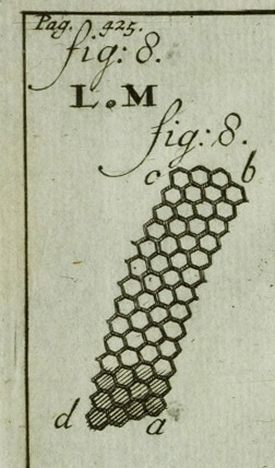
Left: Facets of eye of the adult silk moth. The spot between 'LM' illustrates the actual size of the eye.
Detail from a plate in Leeuwenhoek's
published letters in 'Opera Omnia', vol. 4, 1719. This is an engraving for
the book from the original red chalk drawings by Leeuwenhoek's draughtsman which
is shown on Plate VIII in the 'Collected Letters' vol. 14 (ref.
4).
This engraving has been slightly simplified cf the original chalk drawing which shows a curve of the eye extending from 'bc'.
(The whole plate which includes drawings of wing scales was shared in Part I of this series.)
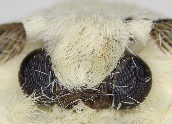
Studying the compound eye of cultivated silk moth Bombyx mori (under stereo microscope and compound microscope)
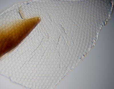
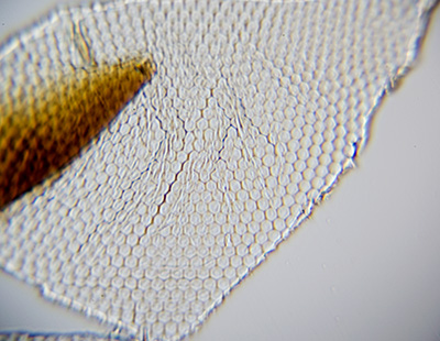
I had a good supply of dead adult moths, courtesy of a silk farm, which offered plenty of opportunity to remove a number of compound eyes and try different preparation and study methods (and lose some!). Although the bodies dry out, the head and eyes seem to show no significant distortion compared with live moths studied. The eyes are quite robust and under the stereo, after clearing the surrounding scales, they were removed fairly readily by slicing with a razor blade behind the eye—although aware
the modern worker, unlike those in the 17th-18th century, has the luxury of binocular stereo, fine razor blades and excellent lighting. Eyes before and after clearing are shown below.
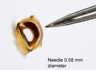
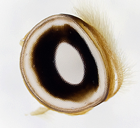
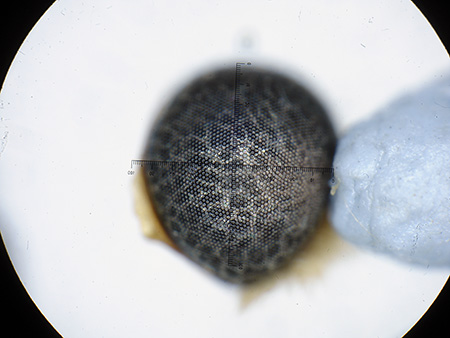
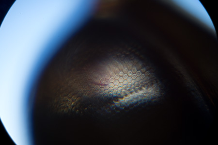
Visual illustration of the typical size of a compound eye (a cleared example in this case, see below) and thus the manipulative skills Leeuwenhoek would require if he counted facets by rotating the eye at a high mag while refocussing.
The eye along widest diameter is typically 1.4 mm.
Clearing in an alkali is a common method for preparing insect facets for prepared slides. I was using long dead dried specimens; Leeuwenhoek's reported method of clearing the eye (of a dragonfly) with a 'fine hair pencil' in water (see below) was likely only suited for freshly killed adult moths.
Optical mag 10X (stereo).
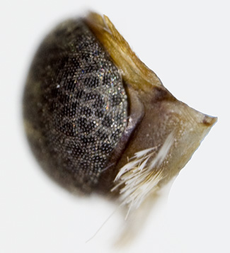
Removed untreated eye. Leeuwenhoek regarded it to be a hemisphere for his facet estimation which it is from the side. I'm uncertain if there are variations, but from above as presented in the insect, the eyes studied to date subtend an angle of ca. 120ş, ie 2/3rds of a hemisphere.
If Leeuwenhoek counted the facets along the quadrant of a whole eye, there seem to be conflicting factors to observation. To help keep track of one line of facets without losing place, a low mag is desirable to maintain depth of field and thus least amount of eye rotation. But the facets are more readily followed along a quadrant at a higher mag, but this is at the penalty of lower depth of field and need for means of accurate rotation with a greater chance of losing
place in count.
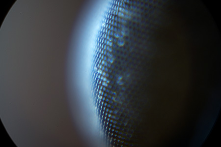
At the optical mag shown 28X (stereo) counting facets along a line seems tricky.
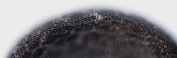
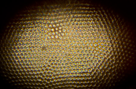
An eye treated in ca. 10% KOH overnight; the facets remain intact. All eye contents clear apart from an opaque annular structure. This ventral view shows that it approximates well to part of a sphere.
Optical mag 20X (stereo).
A modern compound or stereo microscope offers x-y manipulation but not the fine rotational movement required to study the eye facets on the whole eye. (Many earlier pre 20th century compound microscopes did have accessories such as stage forceps / pin offering this type of control.)
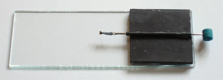
Shown right is a simple homebrew design for rotational studies accurate enough for using a total optical mag up to ca. 100X.
A compound eye from adult Bombyx is shown attached to the needle with BluTak (the needle's 'eye' end for a larger mounting area).
A simple spindle stage. A fine sewing needle has had the smallest grade of electrical heat shrinkable cladding shrunk to the outside, this gives a firm grip along its length and when lubricated with oil allows rotational and x movement. The needle is glued into a notched pad to raise it above the slide. The rubber end allows control.
An untreated eye supported on the 'spindle stage' at the highest optical mag of 80X on my stereo. This shows the curved lenses on each facet. The depth of field is tiny and this mag, for my visual acuity, is about the minimum at which counting facets while rotating the eye was reliable on a modern binocular microscope.
The eye was lit with a powerful LED ringlight under the stereo but the reflectivity of the lenses on untreated eye is low.
Untreated eye under axial epi illumination on the Zeiss Photomicroscope, at a similar mag to the stereo, i.e. total optical mag 75X with 6X objective (with crossed polars for severe flare reduction as not an epi objective).
The axial lighting at this mag is now enough with care to count facets. But it's tricky defining a distinct line of facets on an untreated eye to reliably count along from eye base to apex i.e. along a quadrant without losing place.
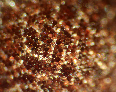
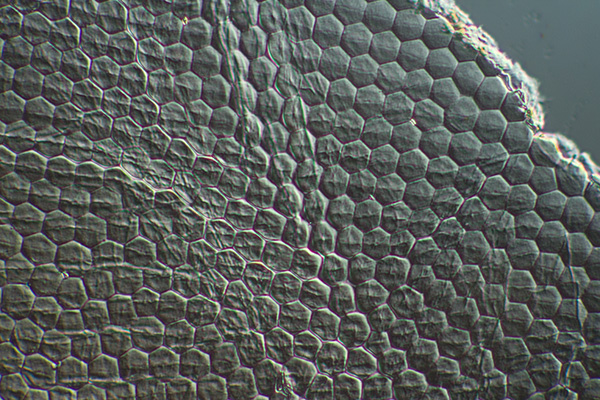
Section of cleared cornea mounted in 50% glycerin in water, Photomicroscope, DIC with Zeiss 16/0.35 objective. A cleared flattened section of facets allows accurate measurement of their size e.g by measuring along a number and averaging.
It is not possible to build a regular polyhedra solely from flat hexagonal facets (a hexagonally tiled planar surface is created—a soccer ball is formed with hexagons and pentagons).
I'm not certain whether the insect compensates by making each facet curved (as distinct from lens surface its attached to), or if it also requires other shaped facets to 'wrap the sphere'. Pentagons and other irregularities are seen but not at high density. A number of pentagons can be seen near the broken edge.
The facets shown are 25.6 µm between the parallel edges.
Studies using a Leeuwenhoek replica
Exploring the subjects Leeuwenhoek studied under a modern microscope is fun and informative but gives no real feel for how he might have prepared, manipulated, lit and studied a subject such as the compound eye of Bombyx under his typical single lens microscopes. To gain a greater understanding of this aspect, its instructive to use a replica microscope. The skilled enthusiast can make either a complete replica (6), or a single lens microscope with homemade lenses in an easier to use mount
(7).
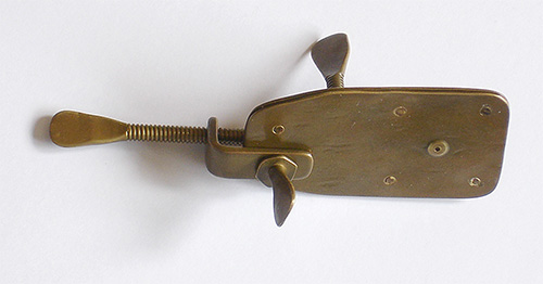
I tried a commercial replica which my brother and I possessed and is shown in use below. It is made by Christopher Allen Replicas, UK.
A modern English made brass replica of a typical Leeuwenhoek design, aged to look authentic and has simply engineered parts in a style typical of Leeuwenhoek's designs.
Shown from the viewer's side, this has to be held very close to the eye. The single lens is shown to the right mounted between two riveted brass plates with dimpled apertures.
This replica is stated to have a ca. 100X magnification (at 250 mm) and was confirmed by my own measurements by projection of a 0.1 mm eyepiece reticle as subject onto a camera sensor. The focal length is ca. 2.5 mm and subject field of view presented to eye is ca. 0.8 mm.
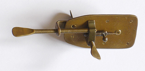
Shown from the subject side, the controls closely recreate Leuwenhoek's designs and allows hands on experience of manipulating subjects.
A pin supports subject and rotates in the 'stage'.
The focus knob (projecting out of screen) acts on the main plate.
The long lefthand screw controls the travel along plate's long axis
The screw just visible behind plate retains the main 'L' shaped bracket and whole unit can swing on this axis. When using the replica I found it convenient to loosen this screw and swing bracket with subject pin fully out to one side to attach subjects.
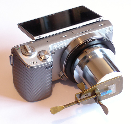
The replica mounted on a Sony NEX 5N digital camera body. Using a C-mount adaptor with card disc to hold microscope in place with BluTak. Additional shaped card formers rapidly centre the microscope lens on camera's optical axis. There are no optical components other than the replica lens.
Note that the adaptor was one to hand, but any lensless mechanical extension suitable for the camera mount can be used e.g. one or more extension tubes. The amount of extension will change the field captured which could be chosen as the full circular field of the lens or a greater extension to crop the field to suit.
The assembly was mounted on a tripod and pointed at either a curtained window (to control light aperture) or an indoors lamp.
The lens to sensor distance was 63 mm i.e. much less than the traditional 250 mm projection distance for formal studies. This projection distance was more practical for the setup and image just filled the APS sensor. This is a similar distance to that adopted by Prof. Brian Ford, a noted authority on Leeuwenhoek's and other makers' single lens microscopes. He describes using 'a lens:image distance
of 60mm' for his impressive 35 mm film photomicrography using the original 'Utrecht' microscope on a sturdy modified 'Leitz micrographic stand' to support
camera and microscope with base illumination (11a).
Leeuwenhoek is known to have used mica plates or thin blown glass to mount aqueous subjects attached to the pin (15) and may have used similar for his silk thread and parts of cleared eye facet studies to keep them flat. I preferred to mount the glass plates using the method shown, where a piece of plastic tube (from Tesco cotton buds) fitted perfectly in screw hole for the pin. With a flat
cut
on end, a coverslip slip fragment
can readily be fitted to face approximately at right angles to lens optical axis.
Hans Loncke shows modified pin designs for his work with the splendid Leeuwenhoek replica he built (6). Perhaps Leeuwenhoek also made alternative supports to the pin for work with flat plates.
The microscope is shown set up to study silk threads from a cocoon mounted in water/glycerin (to avoid rapid evaporation) between two glass slips cut with diamond scribe from a coverslip.
Image right taken using the replica microscope, showing a section of cleared facets from the eye and mounted between two slivers of coverslip. A two foot strip of window light between part closed curtains was used with microscope at a slight angle to the light to give a form of oblique.
The depth of field is small, but although this is a low contrast subject, the facets can be clearly seen, especially along the upper edge; the side lighting gives some modelling. Without using a modern contrast enhancement technique, (e.g. DIC as illustrated above), a modern compound microscope, may not give a superior view to that with a single lens.
An enthusiast like myself is starting from scratch learning how best to use this type of microscope, but Leeuwenhoek would of course developed his skills over many years.
Right, the same subject using the replica microscope with an indoors high intensity fluorescent lamp ca. six inches wide was used i.e. controlling the aperture. A more 'stopped down' look is achieved and illustrates, as with a compound microscope, that controlling the light source aperture has a key role on the image quality and resolution.
Citing Leeuwenhoek's letter dated June 1st 1674, Brian Ford emphasised the importance of controlling the light aperture to optimise the image from a Leeuwenhoek single lens microscope (11b).
Estimating the number of facets and notes on Leeuwenhoek's method Leeuwenhoek's micrometric studies of the adult silk moth's eye are described in his letter dated 20th April 1702 (4). The Hoole translation of the relevant part of this letter is a good read and can be read online at www.archive.org. He counted the number of facets along a quadrant of eye from base to apex and assuming a hemisphere, gave the number of facets along the circumference.
Using a method for calculating the surface area of a sphere described by Metius (17, also see Wikipedia entry for 'quadrature' ) he estimated the number of facets.
After spending some time experimenting with both a compound and stereo microscope, I've had most success to date counting the facets along a quadrant with a stereo at its highest mag of 80X and rotating the eye on the spindle stage described to compensate for the small depth of field.
The image right shows the stereo full field at 80X with widefield eyepieces. The facets are just large enough to follow along a quadrant with rotation but I still found it difficult to keep place; I had to resort to a reticle eyepiece and counted facets as they were rotated through the crosshair.
Strong incident lighting using a ringlight was used.
An untreated whole eye mounted on pin of the replica microscope. Before trying I thought it would not be possible to gain enough oblique light for such a short focal length lens (ca. 2.5 mm) to see facets on the opaque eye. But as the image right shows, with the microscope held at ca. 45ş to the light axis, the lighting is surprisingly good to view the microlenses and doesn't give an image that much inferior to the image captured earlier under a modern stereo
and ringlight, the depth of field looks superior with the single lens.
A line of facets seems more clearly distinguished (and greater possibility of counting them along a quadrant) than the earlier attempt shown using axial lighting on the Photomicroscope.
Shown right is a whole cleared eye under the replica microscope with eye sitting on the subject pin held just by surface tension of the wet eye. The facets are clearly seen.
With this method of mounting the support pin blocks much of the eye so not ideal. If the eye is kept wet to avoid distortion on drying, keying to BluTak fails. Supporting on a tiny glass slip which is then attached to the pin maybe better.
A Sony NEX 5N video of a trial to rotate the cleared eye with minimal changing of focus to count facets under the replica microscope. The full field of view is shown.
If the eye was placed precisely on the same rotational axis as the rotating pin, it seems possible to count facets along a quadrant primarily by pin rotation with a minimal need to refocus.
To rotate along a quadrant from base to apex, as Leeuwenhoek did, requires the eye to be placed with apex of eye facing the lens, i.e just mounted on one edge. For my trial, if the eye was kept damp, surface tension held it to a tiny flat plate mounted on end of pin.
A view through the whole cleared eye under the replica microscope with eye sitting on a coverslip and presented on-axis to the light source. The eye was cleared in alkali which may partially destroy the microlenses on each facet. This may explain my lack of success to date to recreate the multiple images of an object under the replica microscope as Leuwenhoek described.
Creating multiple images under the compound microscope later became a well established 'parlour trick' for prepared sections of cleared insect eyes. Stephanides (14) remarks that to recreate such images the eye should not be cleared 'by long soaking in caustic potash' but cleared with a fine brush (as Leuwenhoek did ) so as not to damage the lenses. But as remarked earlier, fresh eyes may be required for this type of clearing.
Historians have commented that Leeuwenhoek was often reticent to disclose many of his techniques, (e.g. Ford, 16) and it's not clear from his letter of 1702 describing the Bombyx eye studies (4) exactly how he prepared and mounted the eye to count facets along a quadrant. A similar micrometric study of a dragonfly eye in his letter dated 1694 (18) does describe a method of studying a whole cleared eye in some detail
and may also have been used for the Bombyx studies: (Quote from Hoole, 3a.)
'These [the removed dragonfly eye] being placed on a sheet of clean paper, with a small hair pencil and fair rain water, I cleared away the many vessels which fill the inside of the tunica cornea, or horny coat of the eye, leaving only the tunica cornea remaining. This I contrived to place in such a manner, that it might not, as it dried, contract in wrinkles, and placed it before the microscope, and
I often contemplated
it with great admiration, ...'
Immediately after the micrometric studies in the Bombyx letter, he does describe using a cleared eye to use the facets as lenses to view a local church tower. The still images shown above using the replica suggests that a facet count along a quadrant using either a cleared or uncleared whole eye may have been possible by a worker
as skilled as Leeuwenhoek.
I had to use a homebrew spindle stage to mount the eye as fine rotation is not a routine feature on modern microscopes. But Leeuwenhoek's typical microscope design had this feature built in; the pin supporting the subject rotated. Although his microscopes were simply made by modern engineering standards, rotation of ca. 90ş was only required to count along a quadrant and a homemade screw may have been sufficient to
retain good centring while rotating subject.
After trying a Leeuwenhoek replica for the whole eye studies, I did wonder if the modern microscope's wide field optics worked against the user if counting fine repeating features; I needed an eyepiece crosshair with the stereo as a frame of reference. Perhaps the limited field of view together with a small defect in the lens acting as a frame of reference aided such micrometric studies with his microscopes (if so, this may
also
have aided his counting of the fine ribs on the silk moth's wing scales so accurately as described in Part I.)
Two other methods of counting the facets seem possible:
Leeuwenhoek may have cut a part of the cleared 'cornea' and mounted a flattened section in such a way that he knew a quadrant or diameter had been maintained. I did try cutting a cleared eye along the diameter, flattening between glass slips, then counting facets along the cut edge. This did give a count of 40 for half the circumference i.e. a figure close to that for the whole eye studies (see below).
He may also have taken a peel of the 'cornea' and mounted this. He does describe taking peels of the 'cornea' to study the dragonfly eye structure (19). However it would require an undamaged length from a known area of eye to give reliable facet counts along a quadrant. The 'Collected Letters' footnote to this part of the letter remarks that a careful peel can provide either the intact lenses
or an upper layer with a facets imprint (19a). To date, I have been unable to take peels from the long dead adult specimens I possess to try this approach. I'm not sure if taking peels is more readily achieved with freshly killed specimens.
Whatever manipulation and observation method Leeuwenhoek used, he notes in his letter that he counted 36 facets along a quadrant which could be nearly repeated when he handed the microscope to his limner (draughtsman) who counted 35 (the latter value was used for the facet estimation).
Estimates of facet count using various approaches
A summary of estimates of facet counts are given below. John Gorham in 1853 (5) discussed the limitations of Leeuwenhoek's method when applied to an idealised sphere of regular hexagons and proposed that a correction was required.
The number of facets on various insect eyes is widely cited in print and on the web but have struggled to find out if there is a definitive modern way of calculating them from measured parameters. My geometry knowledge is sketchy(!) and thus uncertain of the relative merits of the absolute method used below compared with other approaches. From a practical standpoint it is one of the most straightforward, if valid, on a modern
calibrated microscope. Gorham also proposed other methods for insect compound eyes which I haven't explored; this included punching circles out of the cleared eye and counting the facets either directly or in a shaped rhomb in which hexagons pack better. Other variants of counting the surface area in 'facet units' have also been presented (8) or 'compound eye mapping' (9). My values are higher than Leeuwenhoek's if assuming a hemisphere but become closer when corrected for the eye shape.
All methods are approximate of course, in addition to geometric reasons there's the possible variations in nature which may include variation across specimens, possibly the sexes and generations .... and not least variations between observers!
Source / Method
Measurements made
Facets estimate: for both eyes
Comments
Leeuwenhoek 1702 (refs. 3,4)
35 facets along great circle quadrant
6236
Metius method of quadrature assuming eye a hemisphere.
Leeuwenhoek figure corrected by Gorham (ref. 5)
-
7213
Corrects for packing of hexagons cf squares in a circle.
Present study using Leeuwenhoek method without Gorham correction.
42, 46, 46, 47 counts on different facet lines on long axis of eye. Median 46.
7182
Includes a correction for eye being 2/3rds of a hemisphere.
Present study using absolute measurements of facet size and eye diameter.
Facet width 25.6 µm (average for line of 7). Includes facet wall thickness, see below.
Eye diameter 1.40 mm.
Eye subtends 120ş on one axis.
7233
Facet width gives its surface area. Eye area calculated from eye dimension assuming part of sphere. Eye total area divided by facet area gives facets.
Notes:
The absolute measurements in last row also allow a back calculation of the quadrants expected along circumference. The facet width and eye diameter of eye studied would suggest 43 facets in a quadrant, i.e. within range counted).
In a maths online, it was pointed out that the facet wall thickness needs to be taken into account for greater accuracy, and an equation was provided (20). Although my method of calculating a facet width by averaging along a line of facets did inadvertently account for this.
Leeuwenhoek estimated 90-100 facets along a dragonfly eye's circumference and calculated over 8000 facets. A modern footnote in the 'Collected Letters' remarks that 'This estimate is perfectly correct. Dependent on the species, the larger kinds of dragon-flies have up to 10,000 facets' (18a).
Concluding thoughts
I've certainly greatly enjoyed exploring aspects of the cultivated silk moth and allow a hobbyist like myself to learn more about a fascinating insect in its own right with significant commercial impact, but also a practical insight into the marvelous observational and manipulation skills of the early workers like Leeuwenhoek using single lens microscopes.
Some Leeuwenhoek subjects have been widely reported on (11, 12, 13) but other subjects studied, particularly common invertebrates, don't seem that widely revisited using modern microscope techniques and/or replica singe lens microscopes. There seems plenty of scope (excuse pun) for the hobbyist to explore 'hands on' the less reported subjects coupled with comparisons with Leeuwenhoek's published letter translations and
plates.
Equipment used:
- Manipulation and dissection of Bombyx parts. Meiji EMZ1 10-30X zoom stereo.
- Low power photography, Leica S series 10-80X zoom stereo microscope with trinocular head fitted with Sony NEX 5N body. The electronic first curtain shutter (EFCS) mode ensures no mirror vibration.
- Higher power photography, Zeiss Photomicroscope III, Zeiss optics with Canon 600D body (also EFCS by default). Epi head where required, otherwise transmitted.
Acknowledgements
I would like to thank the following people, but any errors in the above article are solely mine.
- William Hyett of www.silkfarm.co.uk for supplying a number of dead adult moths used in above studies and for earlier studies, the viable cocoons for hatching and for providing valuable advice on incubating them.
- The Science Museum Library (London) staff, in particular Shani Davis, for patiently checking English pagination of Leeuwenhoek letters in the 'Collected Letters' volumes and for copies (via their excellent postal photocopying service). - Research Library staff of the Max Planck Institute for the History of Science for clarifying 'Open Access' image use from the ECHO website (Leeuwenhoek's 'Opera Omnia' bookplates).
- My brother Ian for loan of the Sony NEX 5N digital camera and suggestion to use electrical heat shrink tubing for the spindle stage.
- The Yahoo 'microscopes' forum for valuable discussions on the pros and cons of different approaches to estimate the number of facets on a compound eye.
Notes and references
For a summary of the work of Leeuwenhoek, Malpighi and Swammerdam on the cultivated silk moth see refs. 1 and 1a:
1. M. Cobb, 'Malpighi, Swammerdam and the Colourful Silkworm: Replication and Visual Representation in Early Modern Science', Annals of Science, 2002, 59, pp.111-147.
1a. A. Locy, Malpighi, Swammerdam and Leeuwenhoek', 'The Popular Science Monthly', 1901, vol. LVIII, April, pp.561-584.
2. F. J. Cole, 'Leeuwenhoek's Zoological researches.—Part II. Bibliography and Analytical Index', Annals of Science, 1937, vol. 2, no. 2, pp. 185-235.
2a. F. J. Cole, 'Leeuwenhoek's Zoological researches.—Part I', Annals of Science, 1937, vol. 2, no. 1, pp.1-46.
3. S. Hoole, 'The Select Works of Antony van Leeuwenhoek, Containing His Microscopical Discoveries in Many of the Works of Nature.' 2 Vols, 1798, 1807. The chapter 'On the Silk Worm' is in volume 1 pp. 49-64 and Plate II. The eye studies are discussed on pages 62-63. www.archive.org
has both vols. but plates incomplete. Also available in various modern reprints possibly also without the complete plates if www.archive.org was used as source.
3a. ibid. Volume 2, page 342.
4. 'The Collected letters of Antoni van Leeuwenhoek = Alle de Brieven
van Antoni van Leeuwenhoek', Leeuwenhoek, Antoni van, 1632-1723. Volume XIV, 1996 Swets and Zeitlinger-Lisse. Letter no. 236 [146], 20 April 1702, pp.101-133
(odd numbered pages are in English), and plates VII and VIII. The website www.lensonleeuwenhoek.com summarises this 'monumental' publishing project; vol. 1 was published in 1939, 15 of the planned 19 vols. have now been published (with vols.
16 and 17 due in Dec. 2012 from the current publishers CRC Press). The webpage of theEditor L.C. Palm of the publishing project
tabulates the letters published in each volume to date.
4a. ibid, page 125.
5. J. Gorham, 'Remarks on the Cornea of the Eye in Insects, with reference to certain sources of fallacy in the ordinary method of computing the Microscopical hexagonal Facets of this membrane: with an Appendix, containing a brief note of a new method of taking transparent Casts of the above, and other objects for the Microscope, in Collodion.' Quarterly Journal of Microscopical Science, 1853,
Vol. s1-1, p.76-84. Download free full paper from the 'Journal of Cell Science' archive.
9. Hassan Al Marshad, "A method for compound eye mapping: Dimensions and regional variations in firefly (Coleoptera: Lampyridae) compound eye" (January 1, 2008). Dissertations Collection for University of Connecticut. Paper AAI3308226. http://digitalcommons.uconn.edu/dissertations/AAI3308226
(I don't have access to this reference but the Abstract looks very interesting and describes using a custom made goniometer to study and map the eye with computer mapping.)
10. J. van Zuylen, 'The Microscopes of
Antoni van Leeuwenhoek', J. of Microscopy, 1981, 121/3, pp. 309-328.
(Reprinted in
'Antoni van Leeuwenhoek 1632-1723', Eds. L. C. Palm and H.A.M. Snelders, Rodopi,
Amsterdam, 1982, pp.29-55.) Table 1 summarises the properties of nine remaining
Leeuwenhoek microscopes.
11. Brian J. Ford, 'The Leeuwenhoek Legacy', 1991, Biopress, London.
11a. ibid, p.76ff.
11b. ibid, pp.73-74.
12. A. Schierbeek, 'Measuring the Invisible World', Abelard-Schuman, 1959.
13. C. Dobell, 'Antony van Leeuwenhoek and his "Little Animals"', first published 1932. Dover Edition 1960.
14. T. Stephanides, 'An easy method for demonstrating multiple images in insect eyes', Microscopy (now 'The Quekett Journal of Microscopy'), 1964, vol. 29. pp. 278-279.
15. M. Folkes, 'Some account of Mr. Leeuwenhoek's curious Microscopes, lately presented to the Royal Society, Philosophical Transactions, 1724, XXXII, 446. As quoted by Dobell ref.13, p. 316.
16. B. J. Ford, 'The Van Leeuwenhoek Specimens', Notes and Records of the Royal Society, 1981, 36 (1), 37-59. See p. 45.
17. E. J. Dijksterhuis, 'Mathematics in Leeuwenhoek's Letters', 'Collected Letters, vol. III, 1948, pp. 443-453. (Odd numbered pages are in English. Cited in footnote 30 of ref. 4.)
18. 'Collected Letters', Volume X, 1979, Letter no. 137 [83], 30 April 1694, pp.89-137.
(Specifically pp.125-131, odd numbered pages are in English), and plate VII.
18a. ibid, p.129 and Footnote 44.
19. 'Collected Letters', Volume X, 1979, Letter no. 140 [85], 30 November 1694, pp.153-165.
(Specifically pp.125-131, odd numbered pages are in English), and plates VII.
Revision history:
Published October 14th 2012.
October 15th - minor corrections and revisions.
October 17th - video clip and caption updated. October 19th - section on possible observation methods expanded and a note added on accounting for the thickness of facet walls when estimating number of facets.