|
Diatom arrangements by Johann
Diedrich M—ller (1844 - 1907). |
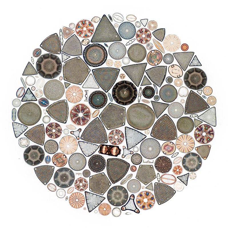
Micscape introduction: Readers familiar with the breathtaking diatom arrangements of Johann Diedrich M—ller (1844 - 1907) may be aware that his most famous arrangement was his "Universum Diatomacearum M—llerianum" of 4026 diatom species. Very few people have had the privilege of exploring this irreplaceable slide under the microscope first hand. In March 2015 Micscape were delighted to hear from a Belgian microscopy enthusiast Jef Schoors who was able to study and photograph the slide using the museum's microscopy facilities and shared an article on this slide and on aspects of M—ller's work. A virtual tour to species level of this slide using Jef's splendid stitched images was also provided.
Jef has recently been allowed access to further slides by M—ller and he shares an image gallery below.
Jef Schoors writes:
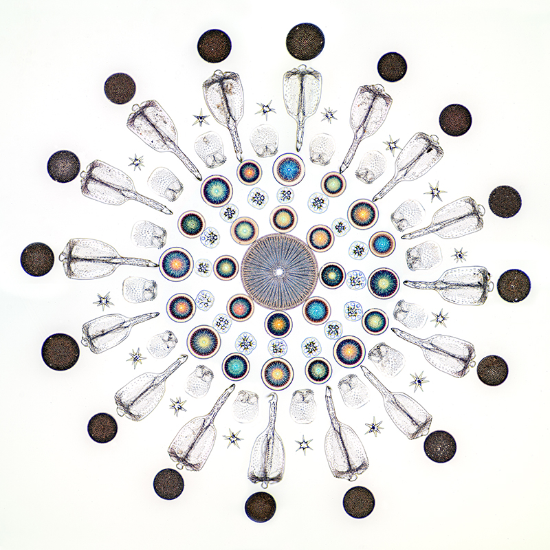
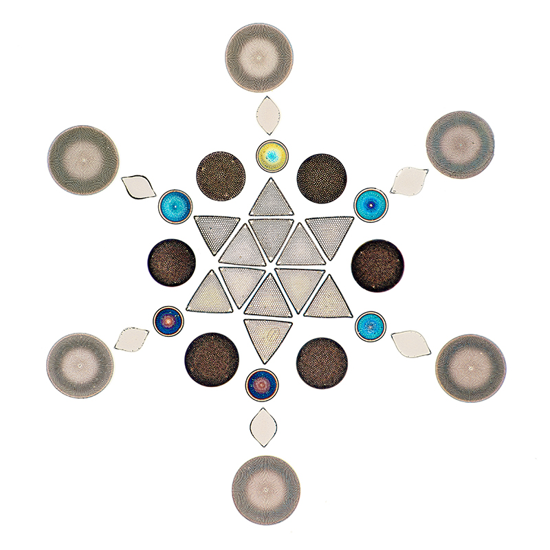
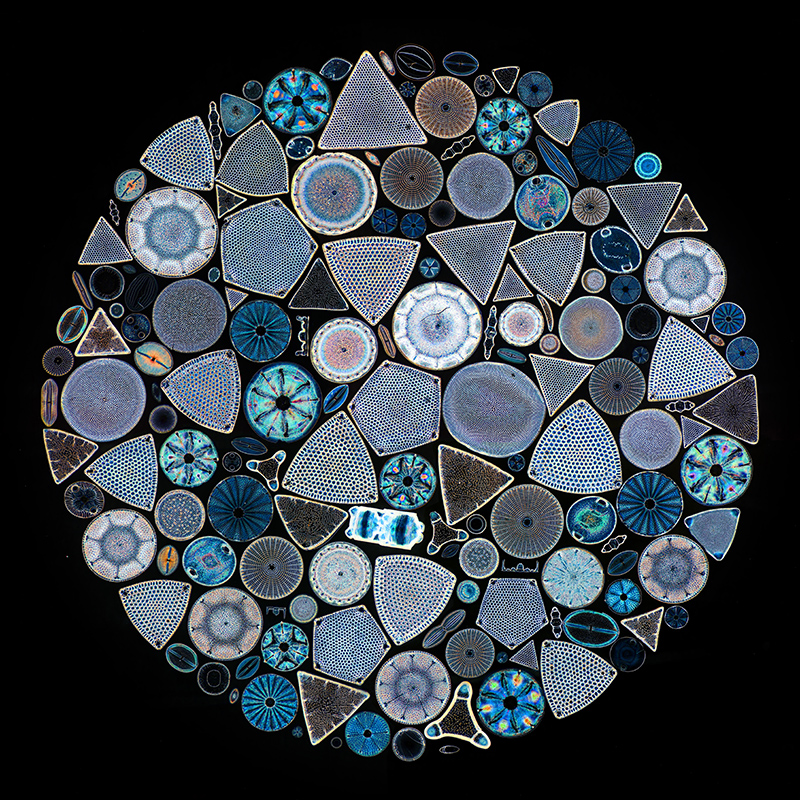
This may be of interest to the visitors of your site: I hereby send you some recent photos that I took of special J.D. M—ller slides which are kept safely in the 'Plantentuin Meise' (Meise Botanic Garden, Belgium). Some of them are also on display in the exhibition 'Diatoms, art in a box of nature'.
The 'M—ller Universum Diatomacearum' was shot in 2015 in the Meise Botanic Garden because this precious slide is stored in a safe and may not leave the laboratory. This also counts for the extensive diatom material and slides from the Van Heurck collection (with lots of M—ller slides).
For the exhibition however I was exceptionally allowed to photograph some diatom mounts with my own equipment at home. Since I always aim for the highest quality I rely on different scopes. The brightfield and darkfield photos are done with my Zeiss Standard and planapo's. For the DIC photos I use a Leitz Aristoplan, equipped with plan Fluotars. The phase contrast photo is done with an old Olympus Vanox and plan phase optics.
I get the best results with the appropriate photo-oculars: Zeiss KPL W 8x/20 (10x would be better because the 8x gives a little vignetting), Leitz Periplan 10x (the one with glasses symbol and the red dot), and Olympus FK 2,5x.
All shots were taken with a Nikon D800 [DSLR] in RAW and cleaned up in Photoshop. I used stitching (2 to 4 images) and to some degree also stacking. For the connection of the Nikon to the microscopes I replaced the original connection rings with Nikon conversion rings (M42 - Nikon) plus some 'DIY adapters' and verified parfocality with the oculars.
The old optics do not have the same clarity as modern equivalents, but the sharpness is there, and the 'haze' is easily filtered out with the RAW convertor of Photoshop.
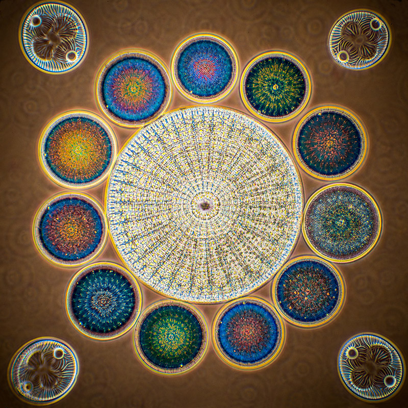
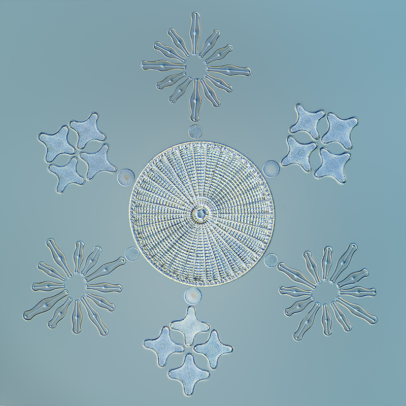
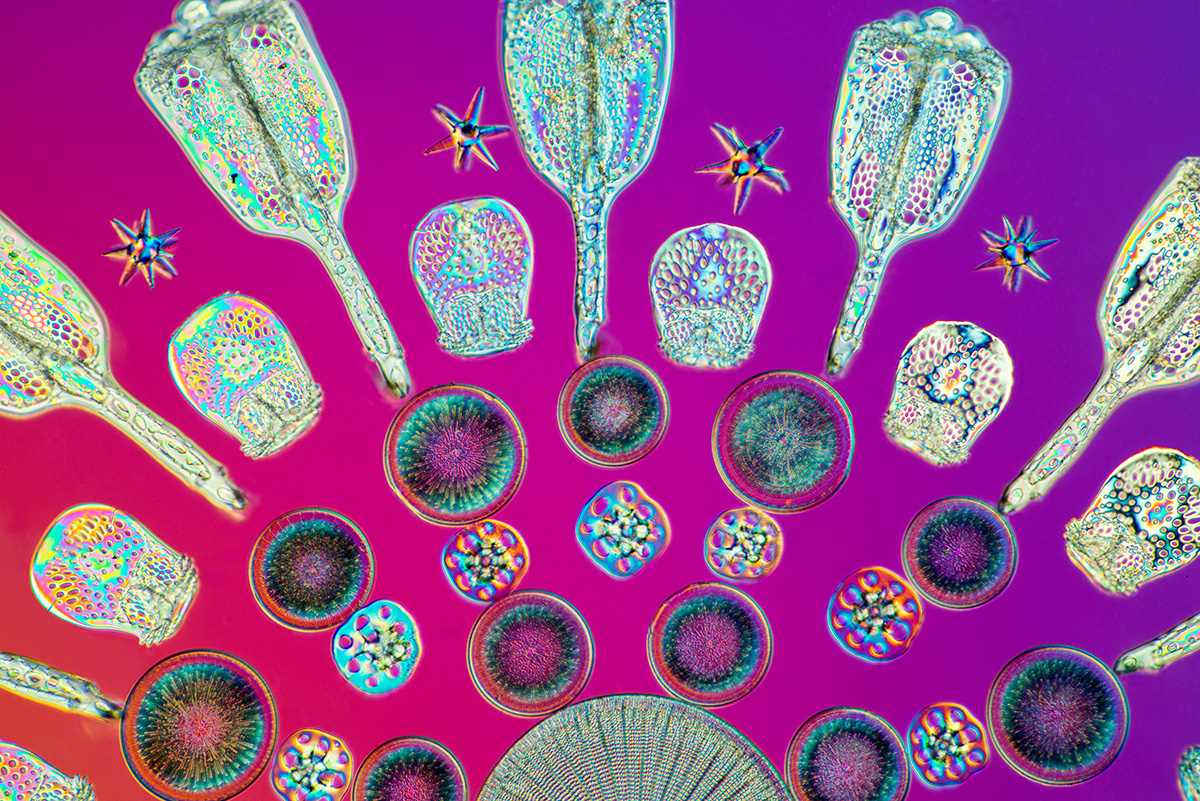
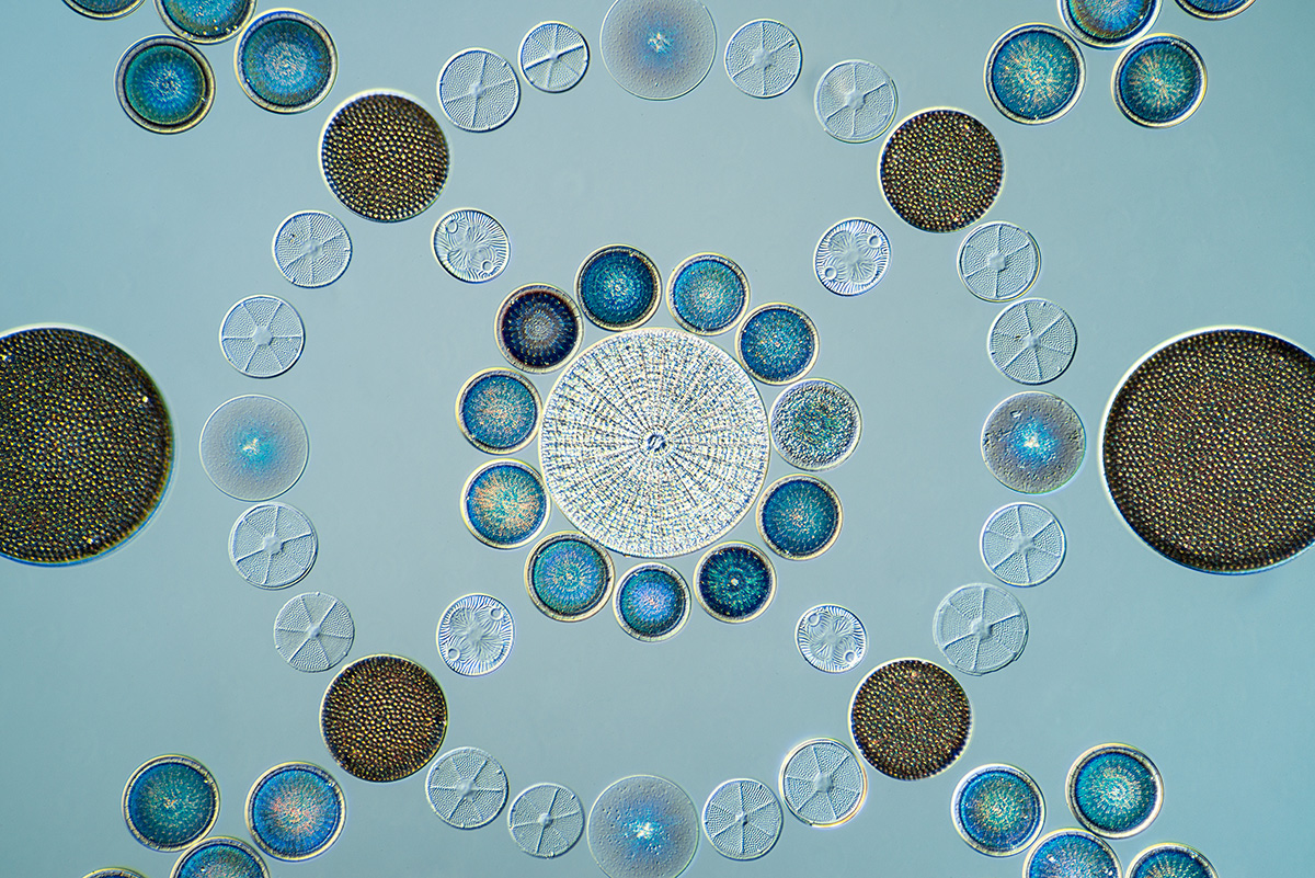
Images - brightfield unless otherwise stated.
Inverted in Photoshop
Phase
DIC
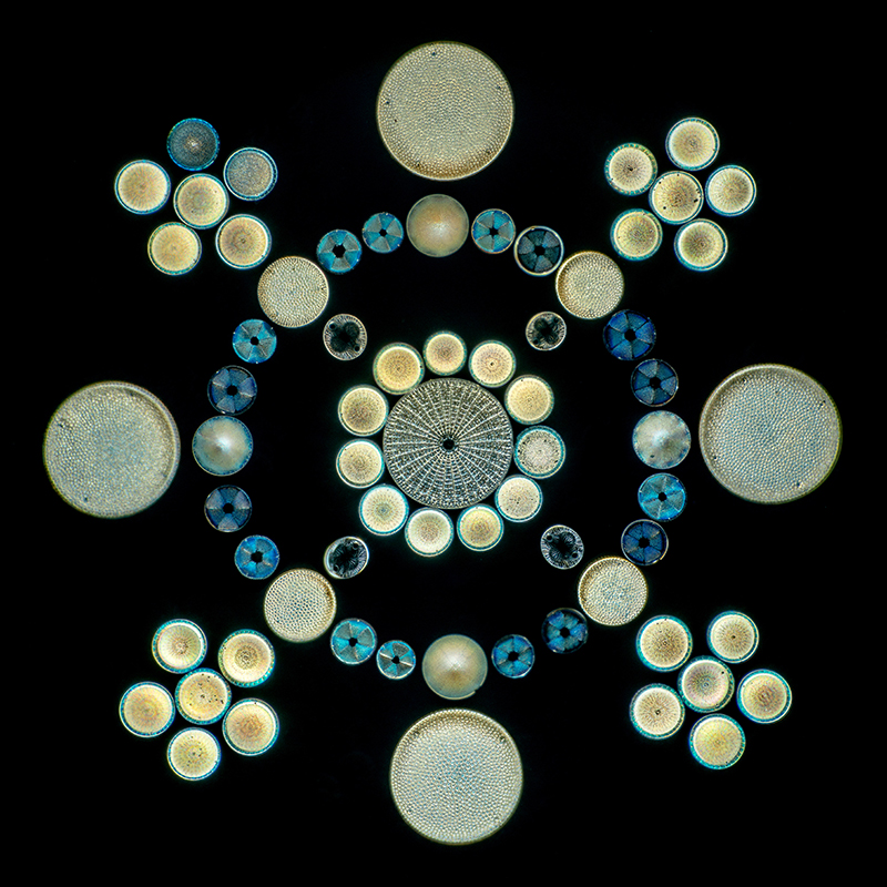
Darkfield
DIC with lambda plate
DIC
Comments to the author Jef Schoors are welcomed.
The images above of the slides at the Meise Botanic Garden are by Jef Schoors. For further image use please contact the author in the first instance.
Selected Resources not linked to in the text:
1) The Van Heurck Collection: A phoenix rises from its ashes., by Bart Van de Vijver, Christine Cocquyt & Jan Rammeloo. Acrobat pdf, copy courtesy of Jozef Schoors.
2) Johann Diedrich M—ller, 1844 - 1907. Die Kunst, Diatomeen zu legen. Ausstellung im Zoologischen und Botanischen Museum der Universitðt Hamburg 15. November 2007 - 15. April 2008. An attractively illustrated article in German on an exhibition in 2008 of his life and work. Acrobat pdf, copy courtesy of Jozef Schoors.
3) Diatomeen im 19. Jahrhundert. Typenplatten und Salonpraparate von Johann Diedrich M—ller by Helene Kranz. Published by Goecke and Evers, 2009. A beautifully presented book in German with stunning photomicrography of many of M—ller's slides by Matthias Burba. Formerly supplied by some dealers but no supplier found either new or used in August 2019. The cover and a selection of pages can still be seen here.
Related Micscape articles:
- Enjoying a M—ller 80 form diatom type-slide with a microphotograph setting by David Walker, Micscape, January 2009. Also has additional references.
- The Micscape Library has extensive resources on many aspects of diatoms by our contributors:
For a splendid introduction see Those Who Live in Glass Houses by Bill Amos, illustrated by Wim van Egmond.
The diatom forms presented on microscope slides are usually the silica frustules (the 'shells'). To admire a selection of live forms visit the 'Micropolitan Museum' by Wim van Egmond and follow the The Marine Collection / Diatom Display.
Published in the August 2019 edition of Micscape.
Please report any Web problems or offer general comments to the Micscape Editor .
Micscape is the on-line monthly magazine of the Microscopy UK web site at Microscopy-UK
ˋ
Onview.net Ltd, Microscopy-UK, and all contributors 1995
onwards. All rights reserved.
Main site is at
www.microscopy-uk.org.uk
with full mirror
at
www.microscopy-uk.net
.