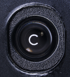
The Foldscope: Exploring the construction of a beta trial kit, the lenses and their optical performance.
by David Walker, UK
The Prakash Lab Foldscope co-invented by Manu Prakash and James Cybulski at Stanford University will be familiar to many
readers. The lab has developed and produced in-house a sub-$1 microscope based on single lenses and folded paper for both educational and potential diagnostic
uses. The project has received very wide publicity and thousands of users
worldwide have being exploring free 'beta trial' examples distributed
from 2014. The Foldscope 'Microcosmos' website for users illustrates the impressive breadth of subjects to which the Foldscopes have been applied to date including by young children, students, hobbyists, teachers and research workers. Both my colleague Mol and myself were very keen to try examples hands-on and signed up for the trials. Unfortunately the kits were lost
in the post but the Prakash Lab have kindly sent us a two microscope kit recently for assessment.
I have a particular interest in single lens microscopes and own two brass replicas based on Van Leeuwenhoek's design, one by Christopher Allen Replicas (UK) (below) and another by Museum Boerhaave (Netherlands), both with biconvex ground lenses (100X
and 85X respectively). I enjoy exploring subjects that Van Leeuwenhoek studied such as the cultivated
silkmoth
Bombyx mori to learn hands-on of his skills. This article explores constructional and optical aspects of the Foldscope.
|
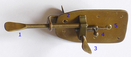
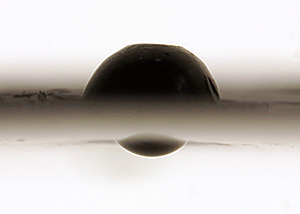
A Van Leeuwenhoek replica microscope made of brass by Chris Kirby of Christopher Allen Replicas (UK). It is typically held vertically against eyebrow ridge (or can be used sideways in eye socket.) Full length as shown 8 cm. The 80X lens is biconvex, ground, focal length ca. 2.5 mm, subject field of view presented to eye is ca. 0.8 mm.
1 - Screw thread allows subject movement along one axis.
2 - If knob on reverse is loosened, the subject can be swung in an arc for the other axis.
3 - Focus knob, the thread acts on the brass plate.
4 - Knob can rotate the subject, also some lateral movement.
5 - Lens secured in dimple between the two riveted plates. Subject mounting pin in front. The pin suits some subjects but pieces of glass or mica were likely used by Van Leeuwenhoek for aqueous mounts.
|
|
The kit and its construction
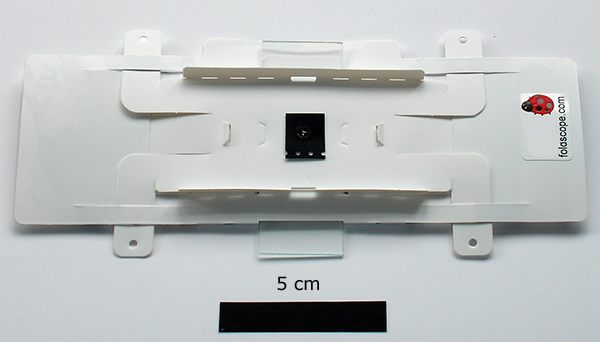
The Prakash Lab have produced an excellent series of four videos on the Foldscope's assembly and use. A typical beta test kit's components are shown below. This example has both the 140X and 430X lens with the LED lighting module. Kits are to be available commercially from August 2017 on completion of the ongoing Kickstarter fundraising campaign with one aim to place 'a microscope in every pocket'. This kit will use
the 140X lens.
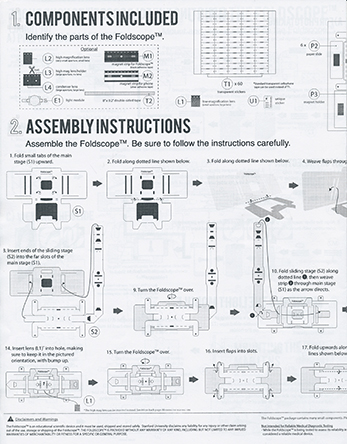
The Foldscope A4 sheet (left above) is made
of plasticised tough paper ca. 0.2 mm thick. The pre-cut components press out easily and includes the microscope 'body', gliding lens / lighting assembly, sub-assemblies for the lens, condenser, lighting unit and six slides. A comprehensive illustrated instruction sheet is enclosed (right above,
half one side). Assembly isn't difficult using the instructions but did find it helpful to watch the videos first and occasionally during construction
to appreciate the procedure better. Once familiar, after completing the first kit, assembly of the second supplied took 10 minutes. The clever design uses solely tabs, folds, slits etc to assemble and create the microscope; double sided sticky tape is supplied to hold firm the lighting sub-assembly and higher mag lens mounts. The Foldscope website shows young children assembling with guidance.
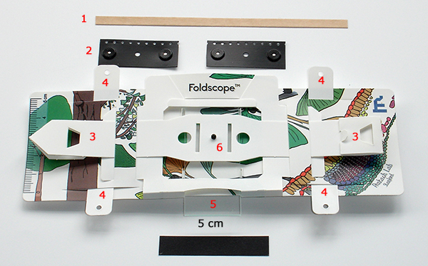
Above. An assembled Foldscope. 1 - double sided tape to secure 430X lens and LED module in their mounts and also for slide preparations, 2 - pair of magnetic holders to align and secure the Foldscope to a suitable smartphone, 3 - tabs flexed with thumbs for lens focus and moved in x-y planes to scan the subject, 4 - tabs that form part of the stage assembly and which
slide in slits which limit the scan motion along the
x axis, 5 - standard microscope slide sits in precut slots, 6 - 140X lens.
Above. Reverse of Foldscope showing the condenser lens in its film mount, push fitted into support (the 140X lens is behind this). The righthand label has a unique
code (blanked out) which an owner can use to subscribe to the Foldscope 'Microcosmos' community website to share project results and participate in forum discussions.
Lighting unit
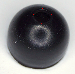
The 140X lens can be used with available light and believe the basic kits widely used in education are of this form. This beta kit was supplied with the attachable LED unit and battery. A ball lens supplied acts as a condenser which is mounted in front of this unit and recommended for use with the 430X lens but can also be used with the 140X lens. The LED module is a neat custom miniature design—small and light enough to be supported by the Foldscope. The objective
lens with aligned lighting module move in unison to scan the sample, in contrast to the moving subject and fixed optics / illumination of a compound / stereo microscope (or Van Leeuwenhoek's design).
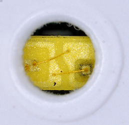
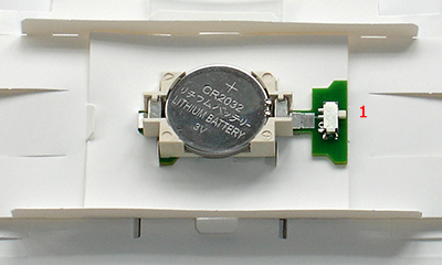
Above. Details of the LED lighting module. Left - the surface mount emitter seen through the ca. 1.1 mm aperture in its card mount. Right - the circuit board mounted in the Foldscope, four tabs hold it secure but can be removed for available light use. It's powered by a replaceable CR2032 battery and operated with switch labelled '1'.
Lens
designs and specifications
The Foldscope uses at present commercial borosilicate spherical ball lenses*—a 140X and in some kits a 430X—their specifications and calculated performance are presented in the 2014 paper by Prakash et al (1). The authors describe the benefits offered by spherical lenses e.g cost, manufacturing, ease of optical alignment but note the future possibility of more complex designs e.g aspheric and/or multi-element
for
superior control of aberrations. Both
lenses are supplied premounted in stiff black plastic film ca. 0.3 mm thick. Given the size of the lenses, the precision and cleanliness to which the lenses have been mounted within the Prakash Lab facilities is impressive even when inspected, as here,
under the stereo microscope. With such small fields of view, the tiniest piece of plastic swarf etc could impair or prevent use. The Prakash paper describes the creation of the precise lens apertures by 'polymer encapsulation' and the 'capillary encapsulation lens mounting' process used. A video of the lab's ingenious 'Foldscope assembly robot' showing lens mounting in action can be seen on YouTube.
*Van Leeuwenhoek's extant microscopes with lenses have ground biconvex lenses except the finest performing (the 266X, 'Utrecht' model, NA 0.37, measured resolution 1.35 µm) which has an aspheric lens believed made by a blown glass technique (see van Zuylen, 1981)(2). Current commercial Van Leeuwenhoek replicas by Christopher Allen or Museum Boerhaave
also
have
ground biconvex lenses as do high quality amateur designs e.g. those made by Hans Loncke (3) and Alvaro de Azevedo (4). The finest resolving single lenses likely made to date are by the noted optical expert, the late Horace Dall, as reported by the late Brian Bracegirdle, these include a microscope with a 430X lens made from yttrium aluminium garnet with NA of 0.6 (5).
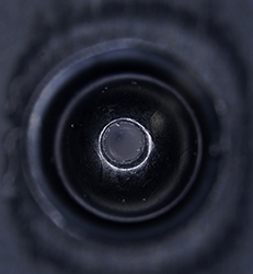
Above. The 140X lens (stated diameter 2.4 mm) in its film mount. The upper lens surface (left) is fully encapsulated within the extruded film and the lower lens
surface (middle) recessed below the plastic film. This should help keep the lens free from dirt as it will sit on a slide surface when not focussed but it reduces the working distance from a covered slide. (The C-shaped light is an artefact from the stereo's LED quadrant lighting.)
The fully encapsulated lens press fits into the card aperture on the lens mount (right). Lens aperture boundary shown in red. Measured lens aperture 0.65 mm (stated 0.7 mm, ref. 1).
Above. The 430X lens in its film mount (stated diameter 0.8 mm). The upper lens surface (left) is fully enclosed and protected within the extruded film. The lower optical surface (middle and right) freely protrudes. This may be necessary because of the small working distance. But in the present Foldscope design it will sit on the subject possibly
uncovered when not focussing and it may be prone to getting dirty, especially in field
use.
Such tiny lenses may be difficult to clean or even to assess if dirty. This may become an increasing problem for the higher mags described which are potentially planned for diagnostic use which may include lenses stated to be up to 2180X with a diameter of 0.2 mm.
This lens is taped onto the underside of a blank larger film plate with extruded hole to fit the Foldscope.
Initial lens trials
I don't own a smartphone with camera built in to securely align and mount the Foldscope with lens, so to initially assess the 140X and 430X lens and share photographs, they were each mounted on a homebrew 'optical bench'. This allowed each lens' capabilities to be explored independently of the Foldscope itself and any limitations e.g. of holding a focus point or subject position, but in due course, the lenses will be assessed on the Foldscope.
Important note on visual cf camera images. A camera field of view may not match the visual field, the latter decreases as the lens is held further from the eye. The camera setup below was capturing ca. twice the visual field when Foldscope held at eyebrow. A photograph also does not necessarily reflect what the user will see from a spherical single lens and visual images in some aspects may
be superior. For example, the image plane is naturally curved which suits the retina better than a flat camera sensor. See Wikipedia entry Petzval field curvature. In a Yahoo Microscope forum discussion in 2015 on 'Foldscope Technical Details', Mervyn Hobden made some very pertinent comments on how the single lens and eye lens should be regarded as a compound system and how this can impact on photographic imagery cf visual. It is well known and also from my own experiences that the
light source and quality can critically affect imagery for single lens microscopes. The built in lamp on the 'optical bench' was convenient but may not reflect visual imagery using the Foldscope's custom LED light and condenser.
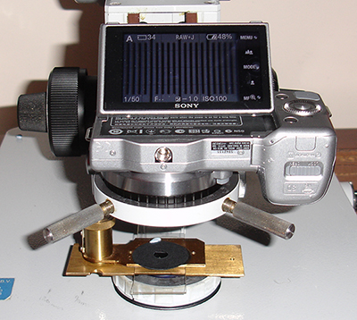
Left above. The 'optical bench' used to take images through the lenses. A Zeiss Photomicroscope with stage, condenser and nosepiece removed allowed the body of a Sony NEX 5N digicam to sit on the condenser mount. The compressorium holding the lens and providing lens focus sat on the field lens mount. Projection distance can be adjusted
using both the condenser mount dovetail and condenser focus. A remote control triggered the camera shutter.
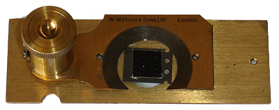
Right above. The lens in its film mount (430X shown) was supported on a piece of clear acetate film with aperture beneath the top plate of a Victorian Watson compressorium. This design has a parallel focussing plate and quite fine focus so provided a stable focus point and subject positioning. The microscope's 100W halogen variable internal lighting was used. (A compressorium was a common microscope accessory to gently compress microorganisms, particularly aquatic, sufficient to immobilise for study without killing
them. Still useful nowadays in the same role.)
Subjects for study could be either temporarily mounted on the glass plate of the compressorium or prepared slides inserted as shown left above. During use, a card stop both on the lamp base and above the compressorium avoided stray light ingress into the camera body and photos were taken with room lighting blanked off.
Lower mag 140X lens
(Stated specs from ref. 1, BK7 borosilicate, spherical, diameter 2.4 mm, source Winston Precision Ball part no. 3200940F1ZZ00A0, back focal length 0.561 mm, resolution - theory USAF 1851 target 1.9 µm, measured 2.19 µm.)
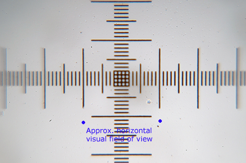
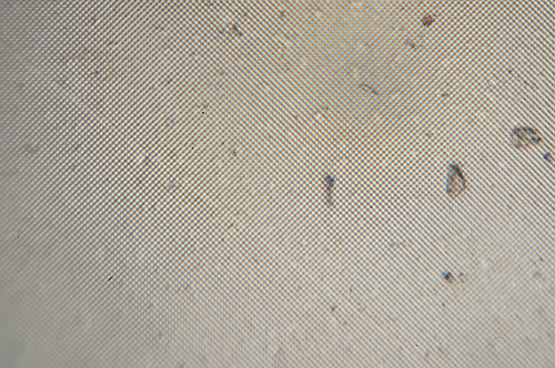
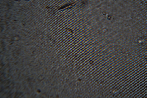
Above. Full camera field of 0.2 mm (Sony NEX 5N with 23.5 mm APS sensor) of an uncovered 0.01 mm micrometer slide, projection distance 68.5 mm. This type of subject shows chromatic aberration well if present but is not marked with this type of lens in ca. 50% of the field which approximates
to visual field. Field flatness good over visual field. Note comment above on the limitation of flat image sensor planes.
The camera field of 0.64 mm gives a value of 134X for the
conventional
250 mm projection distance for citing magnifications i.e. reasonable agreement with the stated lens spec. of 140X.
(Measuring the projection distance to the sensor accurately is not that easy.)
Above. A piece of plastic film two axis diffraction grating marketed by Rainbow Symphony supplied in 35 mm film mounts. Small pieces can be cut out for study. Full camera field of 0.64 mm. The spacing is 200 line per mm using a calibrated compound microscope (although slide
is incorrectly labelled as 13 500 lines per inch or 531 lines per mm.) The lens thus comfortably resolves
5 microns.
An April 2011 Micscape article describes and illustrates the use and calibration of this and the 500 lpm diffraction grating used below.
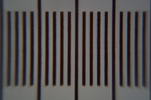
Above. A piece of plastic film of a one axis diffraction grating 500 lines per mm supplied by Rainbow Symphony. Left - full camera field of 0.64 mm, right - detail from same image. The lens thus resolves 2 microns when photomicrograph detail inspected. I could see the resolved detail clearly visually.
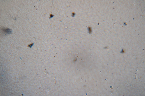
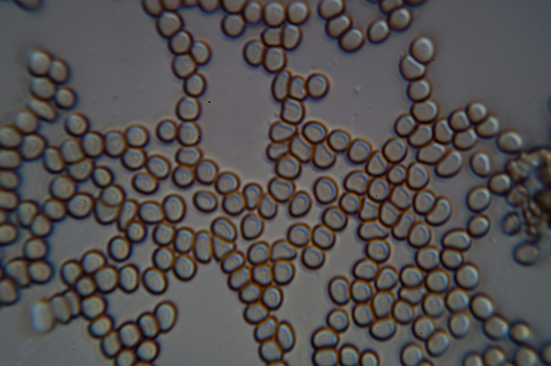
Above. Unstained fresh dried human blood smear. Full camera field
of 0.64 mm.
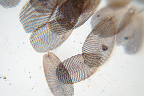
Above. Lepisma saccharina (silverfish) scales. Biosil dry mount with coverslip.
Full camera field
of 0.64 mm.
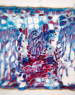
Above. Left. T/S section of the leaf of a sea grape (Coccoloba uvifera). Full vertical camera field, leaf thickness 0.47 mm. Prepared stained slide by Biosil. The visual field was ca. 2/3rds of the stem width
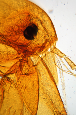
Right. Head of oriental rat flea Xenopsylla cheopsis, a vector of bubonic plague. Prepared slide by John Atkinson (author's brother in law). Field of view vertical, full horizontal camera field 0.64 mm.
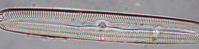
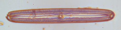
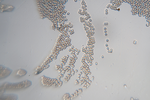
Above. Left - test diatom from a Klaus Kemp 42 form in Zrax mount, taken on a Zeiss microscope with 40/0.75 lens. Right - same diatom using the NEX 5N direct projection 'optical bench', striae unresolved. The dark lines between striae are 1.7 µm apart and were clearly resolved visually with the 140X lens of the Foldscope. Further work is needed to establish what limited this resolution on the camera with the Foldscope.
Chromatic aberration seemed more pronounced for this subject but a green filter did not help.
Higher mag 430X lens
(Stated specs, ref. 1, borosilicate, spherical, diameter 0.8 mm, source possibly Swiss Jewel Co., back focal length 0.187 mm, resolution - theory USAF 1951 target 1.44 µm, measured 1.38 µm.)
The stated back focal length will be very close to the projected image focus distance and is nearly the same as a typical coverslip thickness of 0.17 mm. If a mounted subject is not touching the underside of a coverslip, e.g. with temporary aqueous mounts it may be unable to focus. To date, I've only successfully used this lens on the 'optical bench' with uncovered
subjects. For the present
the subjects studied and photographed below are an uncovered micrometer slide, a fresh dried blood smear and sections from plastic film diffraction gratings.
Above. Full camera field of 0.2 mm (Sony NEX 5N with 23.5 mm APS sensor) of an uncovered 0.01 mm micrometer slide. In the central half of the field, geometric distortion and chromatic / spherical aberration are quite well controlled. The projection distance from lens to sensor plane was the
minimum possible on the 'optical bench' of ca. 63.5 mm. This gives a calculated mag of 448X for the
conventional
250 mm projection distance for citing magnifications i.e. reasonable agreement with the stated lens spec. of 430X.
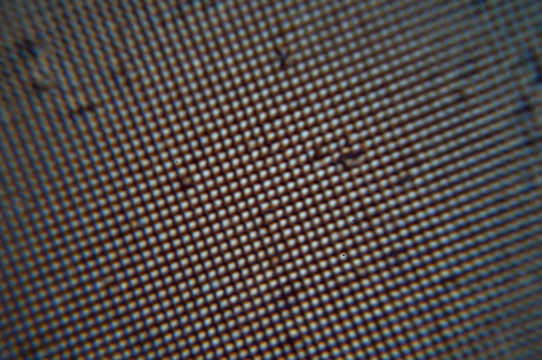
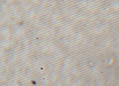
Above. A piece of plastic film two axis diffraction grating marketed by Rainbow Symphony. Full camera field of 0.2 mm. The actual spacing is 200 line per mm using a calibrated compound microscope. The lens thus comfortably resolves
5 microns.
Above. A piece of plastic film of a one axis diffraction grating 500 lines per mm supplied by Rainbow Symphony in 35 mm photo slide mounts. Full camera field of 0.2 mm. The tiny depth of field accentuates any non-planarity of
the film. The lens thus resolves 2 microns. It cannot resolve the equivalent 1 micron
grating and thus its resolution limit lies between 1 - 2 microns. This agrees with its stated spec in the Prakash et al paper (Table 2) of resolving 1.44 microns.
Above. Unstained fresh dried human blood smear. Full camera field of 0.2
mm. The concavities can be seen in some red cells.
Comments
A core aim of the Microscopy-UK website and its ezine Micscape when established in 1995 was to use the then novel medium of the Web to encourage a study of the macro and microscopic world by providing a host for contributors to freely share their skills and enthusiasm online. But my co-founder Mol and I are very aware that our readership base is primarily those who are fortunate to have access to traditional microscopes. A $1 single lens microscope cannot of course
be compared for performance and ease of use with a budget quality student compound microscope (costing from ca. Ł90 in UK) or dedicated field microscope but should rather be viewed for the purposes it was made.
One of the Foldscope project's core aims is to offer affordable microscopes to users worldwide who would not otherwise be able to explore the wonders of the microcosmos. The Foldscope beta trial design for educational
use is ingenious and exploits the performance of the 140X single lens within the limitations of a paper based support. The project members note that aspects of the design are being improved for the next commercial phase i.e a means of locking focus and subject position. The updated LED light source is also noted to act as a 3X/10X low mag lens (Kickstarter
webpage) for initial sample scanning which would be of value to supplement the 140X lens.
The success of the Foldscope concept to date can be judged by the enthusiasm of the users sharing their projects on Microcosmos and the wonder on the faces of many unfamiliar with the microscopic world as they first hold the microscope to their eye. The project to date has distributed 50 000 kits worldwide. The stated aim of the next phase under the commercial arm Foldscope Instruments is to offer a million or more affordable
kits worldwide
in 2017. The Kickstarter campaign to fund it had a target of $50 000 to be raised in 30 days which offers various individual and teacher kits for different amounts pledged. The project leaders note that this target was reached in less than three hours and at the time of writing, pledged funds are over $308 000 with 8 days of fundraising remaining.
Update Dec. 25th 2016. The fundraising finished with 8457 backers pledging a total of $393 357!
It is hoped in due course to adapt suitable projects for the hobbyist currently presented on Micscape for Foldscope users. These will be shared
on the Microcosmos website and on Micscape.
Comments to the
author David
Walker
are welcomed.
Acknowledgement
Thank you to the Prakash Lab, Stanford University for sending a two microscope beta trial kit for assessment to my colleague Maurice (Mol) Smith which is greatly appreciated.
Thank you to Mol Smith for forwarding the kit. A Foldscope with 140X for available light use will be explored in due course by Mol who has a smartphone.
References
1) James S Cybulski, James Clements, Manu Prakash, 'Foldscope: Origami-Based Paper Microscope', PLOS ONE, vol. 9, issue 6, June 2104. (Open access with online additional supporting figures and videos).
2) J. van Zuylen, 'The Microscopes of Antoni van Leeuwenhoek', J. of
Microscopy, 1981, 121/3, pp. 309-328. (Reprinted in 'Antoni van Leeuwenhoek
1632-1723', Eds. L. C. Palm and H.A.M. Snelders, Rodopi, Amsterdam, 1982,
pp.29-55. Thank you to Jan Parmentier for kindly providing a copy of this book.) Table 1 summarises the properties of nine remaining Leeuwenhoek
microscopes.
3) Hans Loncke, 'Making an Antoni van Leeuwenhoek microscope replica', Micscape, July 2007, issue no. 141.
4) Alvaro de Azevedo, a series of articles in Micscape, type 'Azevedo' into the Microscopy-UK 'Find' search engine. Articles include:
'The challenge of grinding lenses for the simgle lens microscope', January 2006 and 'Improving the performance of a single lens microscope', July 2006.
5) B Bracegirdle, 'High Power Simple Microscopes', The Quekett Journal
of Microscopy, 1994, vol, 37, part 4, 296-306.
©
Microscopy UK or their contributors.
Published
in the December 2016 edition of Micscape.
Please
report any Web problems or offer general comments to
the
Micscape
Editor
.
Micscape
is the on-line monthly magazine of the Microscopy UK web
site at
Microscopy-UK
©
Onview.net Ltd, Microscopy-UK, and all contributors 1995
onwards. All rights reserved.
Main site is at
www.microscopy-uk.org.uk
.