|
What is 3D Modelling?
For anyone unfamiliar with 3D Modelling technology, I will provide a short explanation here. A 3D model is a computer-generated
representation of a real object . 3D modellers use software to sculpture, design, and modify geometric shapes to
ultimately create a very realistic model. A texture can be added to the surface of the model in the form of a pre-designed
image, often sourced from real photographic images. Many modern movies are using CGI effects where 3D models are
animated startlingly realistically and interact with real people and world objects seamlessly. A recent film -
the most expensive ever made - called Avatar uses 3D models exclusively instead of real-life actors. The realism
of these models and their behaviour constantly improves annually as the technology increases in its sophistication.
So, what has this to do with Microscopy?
Application in Microscopy,
Biology, and Medicine
When using a microscope
to observe tiny living forms and structures, the observer has to build up a mental picture from what he sees; rarely
can one focus the microscope to see all processes clearly at the same time. Protozoa and similarly sized subjects
are often semi-transparent - frequently possessing rapidly moving cilia, and when alive - can spin, dive, wiggle,
and dart around under the microscope. Larger subjects - flies, mites, and insects - present less of a problem when
viewed through a Stereo Microscope, but can be greater appreciated by young children when seen in a 3D model format,
simply because very young children can find microscopes difficult to use. Another application is where light microscopists
are limited to a scale of about 1600x magnification, and with scanning electron microscopes beyond the reach of
our budgets, 3D models can at least introduce us to the wonders of the bacterial and viral world at the tiniest
scale of living and semi-living entities.
Computers and white board projection screens are fast replacing almost all of the older conventional ways of teaching
in the educational world. The use of 3D models to explain and educate makes it easier to convey initial ideas to
them regarding structure and form of microscopic entities. I don't believe anything can replace individual observation
of real living forms under a real microscope, but I do believe that 3D models can help to encourage further closer
study, especially in the young. This is probably best done by showing static images and video recorded at a microscope
along with the 3D model.
Disappointing
Although thousands of
3D Models can be licenced for a small fee from independent creators via web sites such as Daz3d and Renderosity,
there is a disappointing lack of models created of microscopic or macroscopic entities. Most, if not all, are squarely
aimed at more popular usage. Where appropriate models do exist, they are invariably of insects, with varying quality
regarding realism and movement. I have found almost no models of the type of entity of interest to microscopists:
rotifers, desmids, stentor, etc., One very good web site that licences 3d models for medical teaching is 3DScience.com and I have purchased licences for several of their models.
They have done some remarkable work on modelling the human anatomy but, alas, these models lay far outside of my
modest budget. But their range does include human sperm, ovary, retrovirus, ecoli, HIV, DNA, Neuron, Herpes, Giardia,
Nephron, Avian Flu, Swine Flu H12N1 and Influenza. Some thumbnails of these models can be see in an off-site link here.
Each of them can be licenced for use at a fee of less than $200.
The models are of no use without experience of 3D model posing and rendering software - an application where the
model can be loaded (or many models) animated, lit, and shaded to produce final static images or small videos.
Many of these software applications - Lightwave, 3D Studio Max, Maya - are very expensive for individuals to purchase
but at least two software applications are within our budgets: Poser and Daz 3D Studio (the latter is free!).
Wanted
What would be a great
thing to do, is to encourage the creation of 3D models to cover a wide range of entities we find using a light
microscope and, to this end, I have launched an appeal on here to help raise the money to enable this. As part of this appeal,
I wish to demonstrate the importance and usage of 3D models to Micscape readers and contributors, and Mic-UK visitors,
in teaching and exploring the small-scale world. I have therefore embarked on a series of articles where I will
be using existing 3D models to help greater appreciation of our chosen subject.
The first of these articles covers exploration of the Wasp.
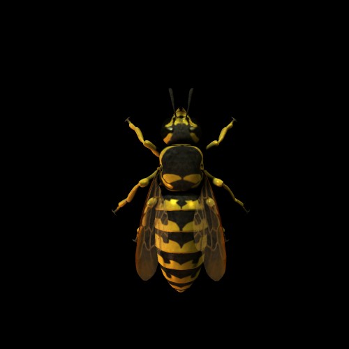
3D model of Wasp
|