|
|
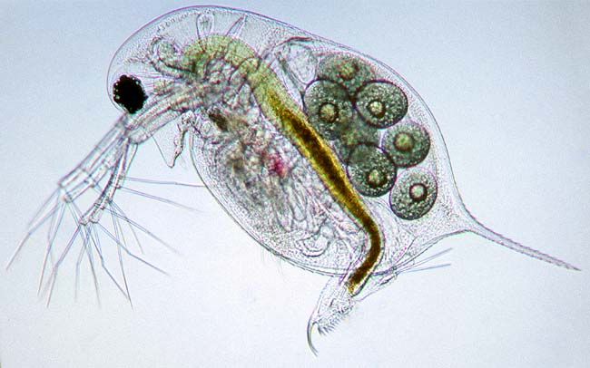
This is a mouse-over image of Daphnia longispina, a common water-flea. Use the underlying image to examine its main anatomical features! |
|
|
|
|
head and eyes |
ovary and embryos |
heart |
Daphnia |
| Water-fleas are easy to find. Most ponds will provide enough of these small crustaceans. They are ideal subjects for study under the microscope. You can examine them swimming under a low power stereo-microscope. When observed with stronger magnification it is best to use a deep slide. You can add dots of vaseline to the corners of the coverslip to prevent the water-flea being damaged. It is wonderful to see the heart beating and the blood flowing! |
All comments to the author Wim van Egmond are welcomed.
Visit Wims home page for links to his many web pages on microscopy
Please report any Web problems or
offer general comments to the Micscape
Editor,
via the contact on current Micscape
Index.
Micscape is the on-line monthly magazine
of the Microscopy UK web
site at Microscopy-UK