Water-flea anatomy - by Wim van Egmond
close-up of the ovary and embryos
b  

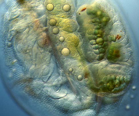
Because of their transparency most small aquatic organisms will give you a magnificent view of many biological processes. Within water-fleas all the stages of the development of the young can be witnessed. In the center of this image we see the long ovary and on the right embryos are developing, already showing red eyespots.

Water fleas don't need a partner to reproduce. By so called parthenogenesis, females can grow enormous amounts of offspring from unfertilized eggs. Male water-fleas only develop when the conditions are getting worse. Fertilized so-called winter-eggs are formed to survive cold but also dry seasons.

This image also shows the skin texture and the gills on the legs.

darkfield Daphnia
brightfield Daphnia
close-up
head and eyes
close up
heart
frontal view
Daphnia


All comments to the author Wim van Egmond are welcomed.

Visit Wims home page for links to his many web pages on microscopy

Microscopy UK Front Page
Micscape Magazine
Article Library


© Microscopy UK or their contributors.

Published in the March 2002 edition of Micscape Magazine.

Please report any Web problems or offer general comments to the Micscape Editor,
via the contact on current Micscape Index.

Micscape is the on-line monthly magazine of the Microscopy UK web
site at Microscopy-UK


© Onview.net Ltd, Microscopy-UK, and all contributors 1995 onwards. All rights reserved. Main site is at www.microscopy-uk.org.uk with full mirror at www.microscopy-uk.net.