Water-flea anatomy - by Wim van Egmond
close-up of the head and eyes
b  

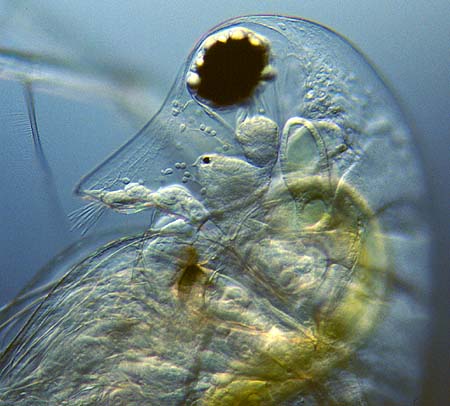
A distinctive feature in water fleas is the compound eye. With higher magnification a second simple eye can be seen as a small black spot. The big compound eye consists of two halves that are fused together. Under the eye the brain is visible. 

The tiny dots left of the brain are blood-cells (corpuscles).

Projecting from the 'snout' (rostrum) are the first antennae with sensory hairs. A digestive gland is connected to the yellow-green gut. 

Five pairs of leaf-like legs create a current in the water that enables them to breath as well as filter food. A pair of pointed appendages pass the food towards two strong mandibles (visible as a brown area).

darkfield Daphnia
brightfield Daphnia
close-up
ovary and embryos
close-up
heart
frontal view
Daphnia


All comments to the author Wim van Egmond are welcomed.

Visit Wims home page for links to his many web pages on microscopy

Microscopy UK Front Page
Micscape Magazine
Article Library


© Microscopy UK or their contributors.

Published in the March 2002 edition of Micscape Magazine.

Please report any Web problems or offer general comments to the Micscape Editor,
via the contact on current Micscape Index.

Micscape is the on-line monthly magazine of the Microscopy UK web
site at Microscopy-UK


© Onview.net Ltd, Microscopy-UK, and all contributors 1995 onwards. All rights reserved. Main site is at www.microscopy-uk.org.uk with full mirror at www.microscopy-uk.net.