Water-flea anatomy - by Wim van Egmond
front view of Daphnia longisipina
b  

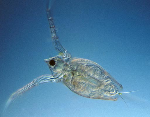
If water-fleas are observed within a shallow dish you can see how they use their powerful second antennae for locomotion. You will also see how feeding takes place using the sieve-like appendages on the legs. These legs are protected by the two halves of the carapace.

This image shows how the two halves of the compound eyes are fused together to form a perfect globe.

Zoom in to see more details

darkfield Daphnia
brightfield Daphnia
close-up
head and eyes
close-up
ovary and embryos
close-up
heart


All comments to the author Wim van Egmond are welcomed.

Visit Wims home page for links to his many web pages on microscopy

Microscopy UK Front Page
Micscape Magazine
Article Library


© Microscopy UK or their contributors.

Published in the March 2002 edition of Micscape Magazine.

Please report any Web problems or offer general comments to the Micscape Editor,
via the contact on current Micscape Index.

Micscape is the on-line monthly magazine of the Microscopy UK web
site at Microscopy-UK


© Onview.net Ltd, Microscopy-UK, and all contributors 1995 onwards. All rights reserved. Main site is at www.microscopy-uk.org.uk with full mirror at www.microscopy-uk.net.