|
Viruses - Twilight Life?
Viruses are not generally considered
to be living entities. They are large molecular units made up of RNA or DNA contained within a protein shell. Unlike
living cells, they lack organelles, ribosomes, cytoplasm, respiratory systems, gas exchange, and a source of energy
creation. They are effectively parasites, capable of reproduction only by hijacking living cells and taking over
control of their reproduction systems, and forcing them to produce more viruses through DNA replication and protein
synthesis.
Viruses for Light Microscopists
Light Microscopists will never see a
virus directly using their microscopes, but it is possible to see cells damaged by viruses in stained sections
of tissue. This means most enthusiast microscopists are limited in what they see for themselves using their instruments.
This is a shame because undoubtedly, most enthusiasts are driven by curiosity and an innate passion to explore
the reality they find themselves in. But we can still explore these twilight entities through 3D modelling and
by using existing data weaned from SEM microscopists and professional study. I suspect we will not be too interested
in the deep workings of the viruses' biochemical processes, or the pathology of the diseases that many of these
viruses cause in plants, animals and humans, so I have kept the content of this article to a style which I hope
will excite your curiosity, and give everyone a 'SEM*1:-less' vision of the sub-light-microscopical world.
SEM*1: Scanning Electron Microscope - wiki
Reasons for the limitations
of Light Microscopes
With a few exceptions (Fluorescence & Confocal Light Microscopy), light/optical microscopes are limited to
the defraction of incoherent light by glass lenses. There is a maximum resolution of all optical systems which
is due to diffraction. The effect is known as diffraction
limitation: the resolution of a
given instrument is proportional to the size of its objective, and inversely proportional to the wavelength of
the light being observed. For a given numerical aperture (NA), the resolution of microscopy for flat objects under
incoherent illumination can be improved using interferometric microscopy.
Several new techniques from emerging technologies will probably break through the current limit of light microscopy
soon by the use of new nano materials to create finer apertures, and the application of coherent light sources
like lasers: the beam of coherent light is different from other light sources emitting incoherent light beams (
tungsten lamps, LEDs ). Our 'normal' light sources are of random phase - varying with time and position; whereas
a laser light is a narrow-wavelength,monochromatic light ( although there are lasers that emit a broad spectrum light at different wavelengths ).
The limitation of an average light microscope ( quality of the instrument may vary this only very slightly ) is
to be able to resolve an entity 0.3 microns*2 across (300nm). Viruses are typically in almost all cases smaller than this. There are
exceptions though! The smallpox virus is exactly this size - 0.3 microns (3 µm*3), and a unique viruses discovered in 1992 is a whopping size at 800 nm (0.8 microns): the MimiVirus!
This is actually visible in a light microscope, although like a star in the sky to an average telescope, it will
be discerned as no more than the tiniest of specks. It is the largest known Virus, larger than some bacterium.
It has been placed in a new family of viruses - the Mimiviridae*x.
*2 A micron, which is a micrometer, is normally stated as µm
*3 Microns to nm (nanometres - english spelling) conversions here
*x Offsite link to viralzone web site
The best way for a light microscopist to understand the size of nearly all viruses, compared to the size of non-viral
microscopic forms, is to present one (a virus) side-by-side to an entity familiar to the microscopist: so, what
about the Euglena? Most of us are familiar with this protozoan*x. Here we are then - a Herpes Virus next
to the Euglena Protozoan, by the application of 3D modelling. The image below shows a slightly exaggerated blue
dot representing the Herpes Virus: it is shown here 4x larger than it really is. Had I shown it at its full size,
it would not be visible at this scale. Please take note that this image, and the movie just below it, may be in
the order of 10x error plus
or minus in what you
see, with respect to the real world, due to issues with presenting accurately sized images in this way on the internet;
a process prone to 'averaging', image pixel issues, and dpi problems. But hopefully it will give you a much better
understanding than by using your imagination alone.
*x Protozoa offsite
link
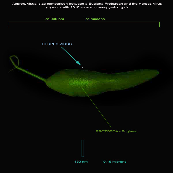
Fig. 1
And since you will have certainly turned up the magnification on your light microscope to take a better look at
Protozoa and other pond-life forms, here is the Herpes virus as it would appear under your 'scope' if you could
see it next to the Euglena and zoom in...
( Note: the accuracy
of scale is + or - x10
)
Note: this is a movie.
Please wait for it to load fully!
So now we have a better understanding of the size and scale of Viruses compared to what we would normally explore
under a microscope, I think it's still frustrating not to see this Virus close up, so let us take a closer look here...
|