An Overview of Viruses for Light Microscopists
A 3D modelling article
by Mol Smith 2010
Please Donate to our Appeal to Fund the Creation of 3D Models for Microscopic Entities!
Please give the pages in this article time to load: medium size video files involved.
You will need a minimum version 6.1 of
Apple Quicktime Player Plug-in to view the movies here.

Page:      1  |  2  |   3   |  4  |  5

  Scale  Herpes Virus  T4 Virus  Influenza Virus   Resources and external links
  To download high res avi files select from here:         Resources and copyrights

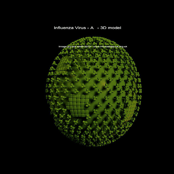
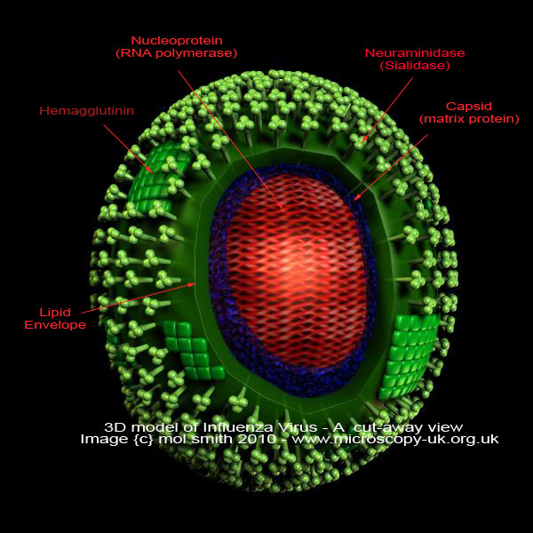
3D Model of Influenza Virus
Influenza virus A is a genus of the Orthomyxoviridae family of viruses. The 'A' type virus consists of only a single species which causes influenza in people, some mammals, and birds. The internal genetic material is RNA, whereas many viruses contain DNA.

The cut-away view below, shows the internal structure and other processes in the virus. Note that is is ovoid in shape and 80 to 120 nanometers in diameter (nm).

Two different varieties of glycoprotein spike are embedded in the envelope. About 80 percent of the spikes are hemagglutinin, which are thought to be involved with the attachment of the virus to a host cell. The other 20 percent or so of the glycoprotein spikes are neuraminidase, which is thought to be part of the mechanism responsible for releasing new virus particles from the host cell. Inside the envelope or capsid is the influenza genome itself, consisting of eight pieces of single-stranded RNA in class A and Class B viruses only.

The animated model below may help provide a better visualization of the Influenza Virus...


You can see a TEM (Transmission Electron Micrograph) image of the Influenza-A virus below. Content Providers(s): CDC/ Dr. Erskine Palmer

Permission
{c} PD-USGov-HHS-CDC (This image is in the public domain and thus free of any copyright restrictions. As a matter of courtesy we request that the content provider be credited and notified in any public or private usage of this image.)


I hope this article has provided a brief insight into the twilight world of Viruses. For a list of resources used in this article and for copyright and licensing information, please see here.

3D Models
A list of 3D models that we are currently aware of as appropriate for biological, microscopical, and medical use is maintained in the 3D model section of our
site here.


Comments to the author
Mol Smith are welcomed.

Microscopy UK Front Page
Micscape Magazine
Article Library

all material © Mol Smith except where indicated


Published in Feb 2010 Micscape Magazine.
Please report any Web problems or offer general comments to the
Micscape Editor.
Micscape is the on-line monthly magazine of the Microscopy UK web site at
Microscopy-UK


© Onview.net Ltd, Microscopy-UK, and all contributors 1995 onwards. All rights reserved. Main site is at www.microscopy-uk.org.uk