|
The T4 Virus
This is often the typical virus shown
in school text books. What a formidable looking structure it has - like something out of a country's military weapon's
system. This is a Bacteriophage Virus - one that invades bacteria rather than animal cells directly - and it one
of a variant of the T4 classification group. This one typically attacks the e-coli bacterium.
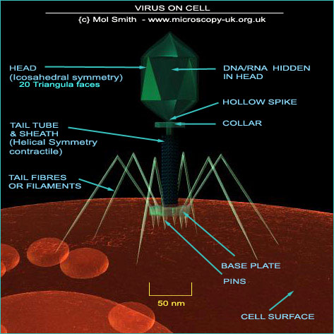
Below is a diagram I made using a 3D model of a T4
virus invading a bacterium cell. The various elements of the structure have been correctly labeled. The internal
DNA/RNA genetic material is not visible in this diagram. Bacteriophages do not just adhere randomly to the host
cell's exterior wall. They 'home-in' on and are attracted to specific receptor sites.
These sites will differ from phage to phage but can
include any part of the bacterium cell, including flagella and pili.
Once the virus has 'landed' on a specific receptor site, all the tail fibres settle down onto the surface and the
base plate become firmly seated on the cell wall, and the tail sheath contracts (the sheath contains ATP WIKI which
is thought to power this contraction). The central tube is pushed through the cell wall membrane and the viral
DNA is extruded from the head down through the tail tube and into the interior of the Bacterium cell. The mechanism
for this action is unknown. The process by which the injected DNA combines with the host cell process is too detailed
and complicated to be explored here, but it is sufficient to say that invasion of a single e-coli bacteriophage
will replicate itself up to 300 times and at a very rapid rate - faster than viruses which infect pant and animal
cells.
I have produced a 3D model animation of a T4 virus locating and attaching to a Bacterium (below), which I have
'artistically positioned' inside a blood vessel. So please note, this is not where e-coli actually are likely to be found: it
just looked cool!
If you look carefully at the approaching T4 Virus, you will see I have made the head transparent, so you can see
the genetic material inside, and its journey down through the viral tube through the bacterium cell wall and into
the interior.
Perhaps one of the most common viruses affecting us humans and causing annual misery is the Influenza Virus, which we can take a quick look at here...
|