An Overview of Human Cells for Light Microscopists
Part II - Human Skin & Human Lungs
A 3D modelling article
by Mol Smith 2010
Please Donate to our Appeal to Fund the Creation of 3D Models for Microscopic Entities!
Please give the pages in this article time to load: medium size video files involved.

Page:      1  |  2  |   3   |  4  |                        For Part 1 The Human Cell - go here!

 Skin  Lungs1  Lungs 2    Resources and external links
  To download high res avi files select from here:          Resources and copyrights
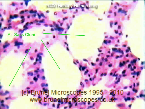
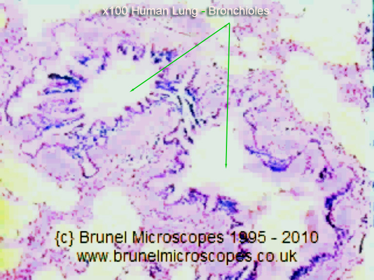
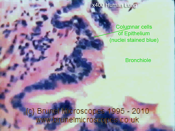
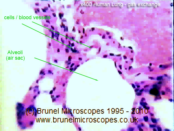
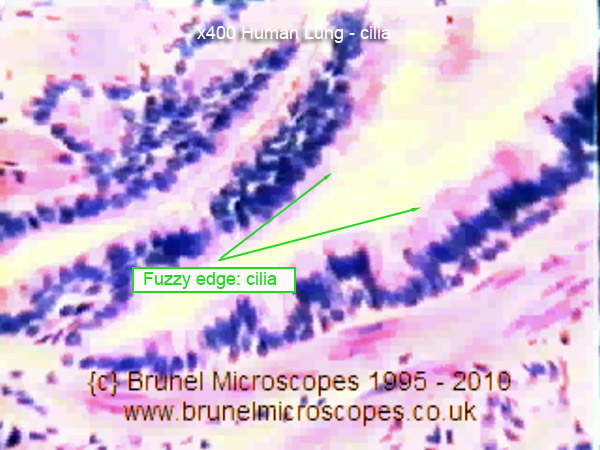
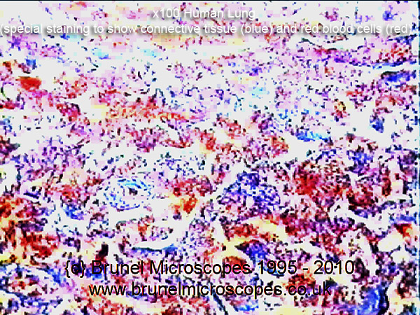
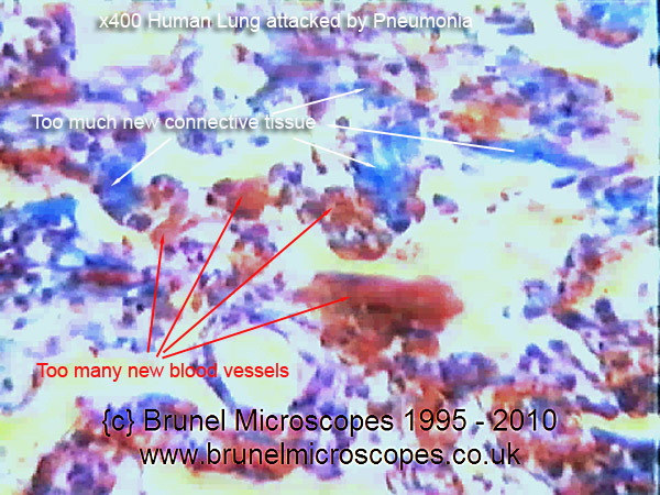
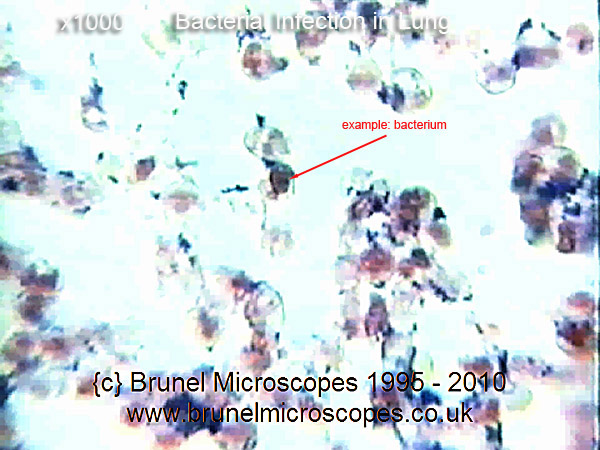
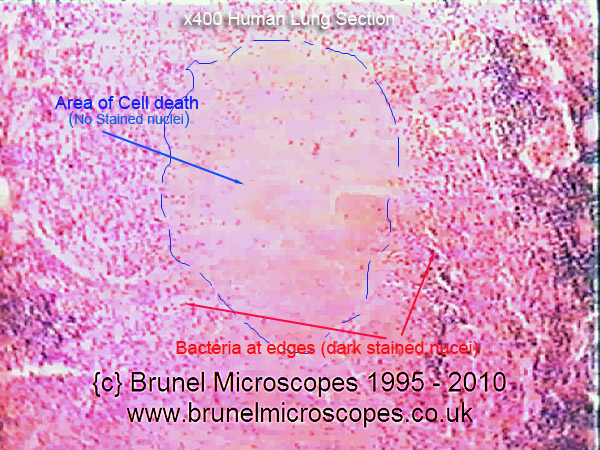
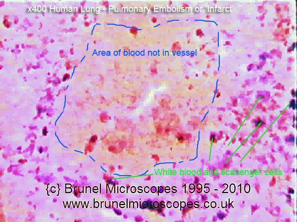
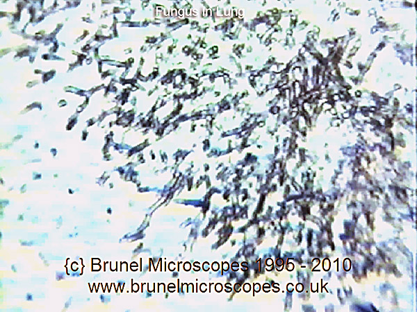
Lung Disease
As with any subject studied under an optical microscope, what you see and determine correctly depends on several things. The first is understanding what staining technique was used in the specimen, as different techniques can reveal different structure and processes. The second, is often understanding what a specimen looks like when healthy and undamaged, and the third is knowing something about interpreting flat 2 dimensional patterns and detail into a 3 dimensional comprehension. Is this circular dot like that because of the way it was sliced? Is that curly thing with the thick blue perimeter really a blood vessel, etc?

As we are now going to look at thin sections from the human lung, I think it is also a great opportunity to develop these skills a bit further. So I will include some advice concerning interpreting staining results and how to recognise structures in thin sections here. Don't think I am an expert at this, because I am not even a very good beginner: I simply listened to the Brunel Microscopes Video and I have used their knowledge for what follows (used with kind permission - see
resources page). The follow images were taken as stills, exported from a VHS video tape (a copy of a copy) so please excuse the image quality. They are fit for purpose in this article though. To see a larger version of any example below, simply click on the existing small image.

         
x 100 Healthy Lung   Healthy Lung - clear air sacs   x400 Healthy Lung
  These lung sections are stained with Haematoxylin wiki and Eosin wiki which will stain cell nuclei blue and the cell Cytoplasm wiki pink. The healthy human lung, seen under a microscope, will show typically clear air sacs (Aveoli wiki). These will be surrounded by cells of the lung comprising small capillary blood vessels, bronchioles, and the Aveoli. The two slides (left x100) and (right x400) here can help you understand this complex network, which is required for gas exchanges to take place.  
 x100 Healthy Bronchiole   Healthy Lung - clear Bronchioles     x400 Healthy Bronchiole
  The Bronchioles (Bronchiola) can be seen here in cross-section as large irregular empty spaces surrounded by the dark blue nuclei of Epithelium wiki cells which represent the Bronchiole Wall. In the x400 slide (right) it is possible to see these cells are columnar in shape with the nuclei at their base and the remainder of the cell cytoplasm extending away from it. Some of the cells may appear less columnar, depending on their orientation, and where they were sliced in the thin section.  
x 100 Gas Exchange   Healthy Lung - Gas Exchange and cleaning   x 400 Cilia
   Gas exchange takes place in the Aveoli (tiny air sacs) through the network of fine blood vessels (capillaries) surrounding each air sac. Oxygen passes into the capillary, and carbon dioxide along with other waste gases are passed back into the air sac to be exhaled. In a healthy lung, the cells lining the Bronchioles and air sacs are lined with fine cilia which 'waft' or sweep any mucus back out and upwards away from the lung processes. They show up as fuzzy edges on the pink cell cytoplasm (see right)
In diseased lungs, the cilia are destroyed, and mucus is cleared only by the action of coughing!
 
x 100 Pneumonia Infection   Diseased Lung - Pneumonia   x 400 Pneumonia Infection
  The specimens both left and right here have been stained using a method called NSB Trichrome (off site), which shows connective tissue as blue, and blood vessels and blood cells as red/orange. Here, too much connective tissue and new blood vessels have formed as part of the body's (lung) defensive strategy to fight infection (get more white blood cells in!). This severely interferes with gas exchange in the lung. The lung sections shown left and right illustrate a lung infected with Pneumonia. Most changes to the lung are reversed once the infection has been medically treated.  
x 1000 Bacterial Infection   Diseased Lung - Bacterial & TB Infection   x 400 TB Infection
  These slides are stained with Haematoxylin and Eosin.
Right slide shows area of
Caseous Necrosis wiki
(cell death) where large areas of cells lack the blue stained nuclei. This has been caused by an infection of Tuberculosis wiki bacteria which can be seen at the edge of the damage. The bacteria causing TB are difficult to treat even with antibiotics.
The slide left shows gram positive stained cocci -{spherical} bacteria infecting the lung. these are relatively easy to clear using standard antibiotics.
See
Staining Techniques wiki regarding Gram Staining.
 
x 400 Lung Infarction   Diseased Lung - Infarct and Miner's Lung   x 100 Miner's Lung
    The slide left shows large areas of lung tissue filled with red blood cells not contained in blood vessels. They are surrounded by wandering scavenger cells and new connective tissue trying to repair the damage. Where an area of lung loses its normal blood supply and starts to do die, the process is called an Infarct wk. In time, the lung may repair itself by removing the dead cells. But not so in the slide of Miner's Lung (right)
where carbon (soot/coal) granules have filled lung structure and alveoli with deposits. The lung tries to clear the damage with white blood cells, which fail and die in situ, increasing the area of non-functioning lung. Smoker's lungs are also irreparably damaged this way.
 
x? Fungus in Lung   Diseased Lung - Infarct and Miner's Lung   Movie of Lung Histology
    Fungal infection in lung (typically Farmer's Lung wiki) caused by spores from damp hay. Hyphae wiki can be seen as rosette clusters of stranded dark (Metallic Stained) filaments. These infections are treatable.

If you click on the image to the right, you will be able to view a very low resolution version of a section of film about the Lung extracted from
Brunel Microscopes Histology Video.


Next month, I will continue this series by looking at other cells in the human anatomy with the aid of 3D virtual modelling.
For credits, resources, and permissions,
please go here.


Comments to the author
Mol Smith are welcomed.

Microscopy UK Front Page
Micscape Magazine
Article Library

all material © Mol Smith except where indicated


Published in May 2010 Micscape Magazine.
Please report any Web problems or offer general comments to the
Micscape Editor.
Micscape is the on-line monthly magazine of the Microscopy UK web site at
Microscopy-UK


© Onview.net Ltd, Microscopy-UK, and all contributors 1995 onwards. All rights reserved. Main site is at www.microscopy-uk.org.uk