|
Tough teeth - the radula of the common limpet
by David Walker,
UK
A look at a classic type of microscope subject, prompted by scientists' studies in 2015 where the teeth were found to be the strongest biological material tested to date.
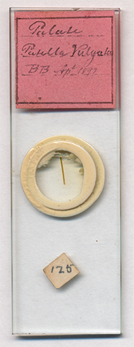
Image right, Victorian prepared microscope slide,
labelled 'Palate Patella Vulgata BB Apr 1897'.
Updated September 29th 2019 with a note on the likely mounter
kindly provided by Brian Stevenson.
|
|
It's not often that the finer points of invertebrate anatomy reach the news headlines, but the teeth which form part of the radula of the common limpet Patella vulgata achieved just that in February 2015. This humble little critter which lives on rocks found along the seashore quietly munching plant matter using its radula to scrape the hard surfaces had been harbouring a secret. Its teeth were made of an organic-mineral composite that has been found to be the toughest biological
material tested to date, demoting the widely cited spider silk into second place.
As a microscopy enthusiast I was delighted because snail radula are one of my most favourite subjects to study—they respond well to a wide variety of microscopy techniques, both incident and transmitted, including autofluorescence for some dry mounted species. After reading the news story I browsed my prepared slides of radula to find that I didn't have an example of the limpet (no shortage
of whelks though). Fortunately, keeping an eye on eBay I found an example for a very modest price—the limpet's newly gained celebrity status didn't prompt a buying frenzy.
The slide is shown above. If 'BB' are the mounter's initials, they are not familiar to me and have found no reference to them, but the seller offered some valuable provenance in the auction (with thanks) suggesting it was a B. Borrows of Darlington (UK):
"From a large collection from the late Dr Black of Seaton Delaval
(Northumberland). A lot seem to be in the hand of R Borrows and his father B Borrows of
Darlington Co Durham and the vast majority date from the 1870's to 1900 with the
latest dated one I have found so far at 1921."
Updated September 29th 2019: Thank you to Brian Stevenson who maintains a splendid resource microscopist.net on slide mounters and has just finished an article on "Richard Borrows, 1864 - 1945". He writes "You speculated that the letters "B.B." on your slide may have been the father of Richard Borrows. Richard's father's name was George. Most likely, "B.B." indicates that Borrows used benzole balsam as the mountant."
The slide hasn't been made to the highest standards, the radula has been lain out linearly as is typical but a number of the teeth seem damaged (in life or during the preparation?) and the white sealant has encroached into the mount. But it provided a good enough example to explore.
The teeth of many radula are opaque so a mix of transmitted and incident lighting is a useful method to provide some detail on the teeth as well as of the underlying structure. I do have the proper incident lighting head for my Zeiss Photomicroscope but rarely use it for covered subjects—I find the on-axis lighting rarely suited for the subjects I like studying. For very little cost and more effective for many subjects,
is off-axis lighting using for example a white LED on flexible neck. I changed out the 5 mm LED on a commercial example for a 3 mm so can shine light at close quarters using objectives with short working distances. I was first alerted to the commercial USB LEDs on goosenecks from the Micscape March 2012 article by Andy Chick who was in turn inspired by Andrew Entwistle's LED lighting project presented in Micscape June 2006.
The two images below show off-axis lighting using the LED combined with transmitted light, firstly DIC, then phase.
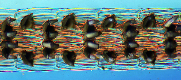
Off-axis incident lighting + transmitted DIC, Zeiss 6.3X NA0.16 with lambda tint plate. Stack of three images.
(Not true DIC as don't have the correct matching prism for the 6.3X, the number II prism with condenser lens off gives reasonable results albeit without an even background.)
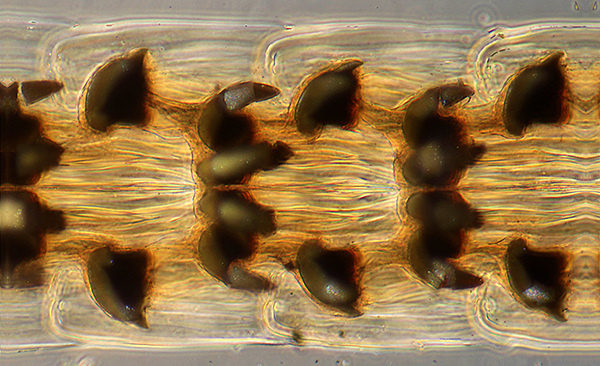
Off-axis incident lighting + transmitted phase, Zeiss 10X NA0.22 Ph objective. Stack of three images.
This example had been mounted in Canada balsam so it was not possible to see if it autofluoresced (the balsam's own autofluorescence swamps that of the subject). See the Micscape Resources below for an illustrated article on examples that do. The fluorescence can be bright enough to allow images to be captured under the low numerical apertures of a stereo microscope.
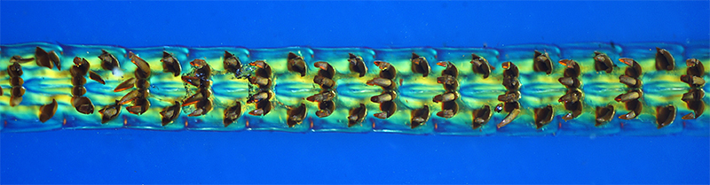
Stereo microscope image, Leica S8 at 5X mag setting. Transmitted crossed polars with lambda tint plate plus incident fill-in light from white LED to give some detail in the teeth.
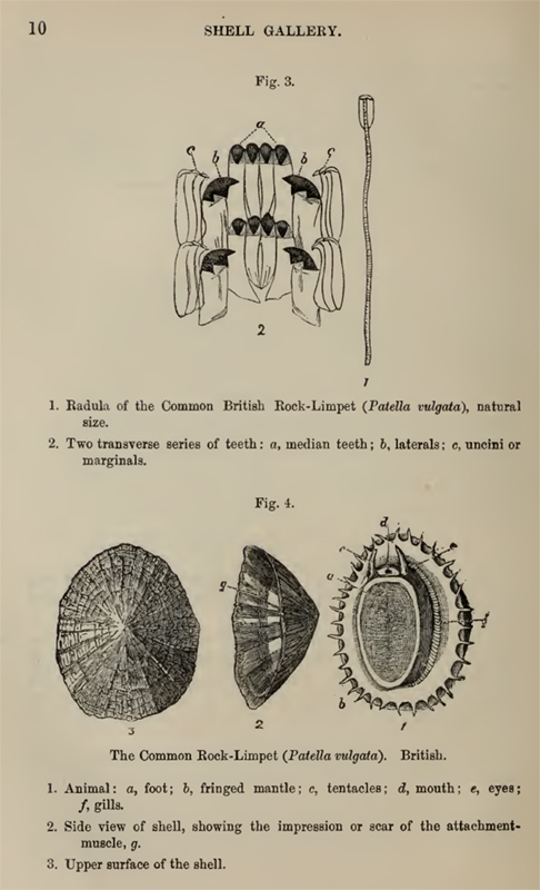
A splendid illustration shown right illustrates the teeth layout and the limpet anatomy. This has been taken from 'A Guide to the Shell and Starfish Galleries' published by the Department of Zoology, British Museum (Natural History) 1901. Preface by E Ray Lankester. (In the public domain at www.archive.org.)
The accompanying text (my underlining and spacing) notes that:
Both the true and the false Limpets are littoral and found on rocks between tide-marks. They have the power of excavating the surface to which they attach themselves, and adhere so firmly that it is easier to break the shell than detach the animal.
The largest known Limpet (Patella (Ancistromesus) mexicana, case 7)
inhabits the west coast of Central America, its shell having sometimes a length of 12 inches. The Limpets are vegetable feeders and fond of seaweeds of various kinds, which they rasp with their remarkable spiny tongues.
That of the common English Limpet (P. vulgata, Fig. 3) is longer than the shell itself, and armed with as many as 1920 glassy hooks in 160 rows of twelve teeth each.
The Limpet is commonly used for bait in the sea-fishing off the Scottish coast, and vast quantities are consumed as food in some parts of Ireland.
Some Limpets, such as P. compressa, P. mytilina, etc., are found on the stems of floating seaweeds, and have the shells usually thinner and smoother than the Rock-Limpets, which have to resist the fury of the breaking waves.
The recent work by Asa H. Barber , Dun Lu and Nicola M. Pugno
On February 18th 2015 the humble limpet and its dental work hit the news headlines because a paper published by the above workers on the same day reported their studies of the limpet radula. They had shown that its teeth are the strongest biological material tested to date. This paper is in the public domain on the Royal Society website (link below). Popular articles were also published e.g. those listed below in Resources and are a good starting point to read of their work in brief.
The image sequence below, shared with thanks courtesy of the Royal Society Creative Commons Licence, shows the clever techniques which they used to prepare a 'dog-bone' beam from a tooth for the mechanical testing. A focused ion beam (FIB) was used to shape it and an atomic force microscope (AFM) to test its mechanical properties. Analytical studies of the tooth construction revealed why the teeth were so strong—they are made of a composite of organics and the
mineral Goethite (an iron oxyhydroxide FeO(OH) ), see Wikipedia Goethite entry. The shape and size of the mineral fibres was a key aspect to minimise flaws in the matrix which may otherwise weaken the structure.
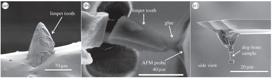
Above. Figure 3 from the reference below. Caption reads "Scanning electron micrographs showing (a) the limpet tooth prior to FIB
milling, (b) FIB sectioning and attachment of the limpet tooth cusp to an AFM
probe and (c) further FIB milling to thin the sample towards a ‘dog-bone’
geometry."
"© 2015 Asa H. Barber , Dun Lu , Nicola M. Pugno. Published by the Royal Society under the terms of the Creative Commons Attribution License,
which permits unrestricted use, provided the original author and source are
credited."
Source: Extreme strength observed in limpet teeth Asa H. Barber, Dun Lu , Nicola M. Pugno, Interface (Royal Society Publishing), volume 12, issue 105, April 2015 (published 18th February 2015). Link is to full free access with pdf download option.
An insight into how nature has developed such strong materials can provide inspiration into how man can mimic these materials for our own use. Nature once again leads the way!
Comments to the author David Walker are welcomed.
Micscape resources on snail radulae
A "palette of palates". Exploring a selection of snail radula - a classic type of Victorian microscope slide David
Walker
Forays into Fluorescence - 4. Notes on the autofluorescence of old dry mounts
of snail radulae. David Walker
Trials of studying autofluorescence with a stereo microscope
using blue or near UV LED excitation David Walker
Snail's Teeth, Spicules, and Other Bizarre Delights: Or Beauty is in the Eye of the Beholder Richard Howey
External Resources
Limpet teeth set new strength record Children's BBC website with a neat video summary, 18th February 2015.
Limpet teeth set new strength record BBC News website more detailed article, 18th February 2015.
©
Microscopy UK or their contributors.
Published
in the May 2015 edition of Micscape.
Please
report any Web problems or offer general comments to the
Micscape
Editor .
Micscape
is the on-line monthly magazine of the Microscopy UK web site at
Microscopy-UK
© Onview.net
Ltd, Microscopy-UK, and all contributors 1995 onwards. All rights
reserved.
Main site is at www.microscopy-uk.org.uk
with full mirror at
www.microscopy-uk.net
.