| | |
| | by Wim van Egmond |
| What are diatoms? |
Diatoms are delicate unicellular organisms that have a yellow-brown chloroplast that enables them to photosynthesize. Their cell walls are made of silica almost like a glass house. The construction of the cell wall, called the frustule, consists of two valves that fit into each other like a little pill box. The colour of the chloroplast is yellow-brown instead of the green we know of other creatures that use light as a source for energy. There are two different groups of diatoms, the pennates which are pen-shaped and the centric which are like a cylinder. In fresh water most diatoms you will see are of the pennate type. | |
| Where
can you find diatoms? At the end of the winter they are most numerous in fresh water. They will cover surfaces of aquatic plants or poles and wooden borders of ponds. If you like to study them you can scrape the brown growth with a flat piece of plastic. You can also use a sponge. For the free living (plankton) species a special fine mesh plankton net is very useful. | |
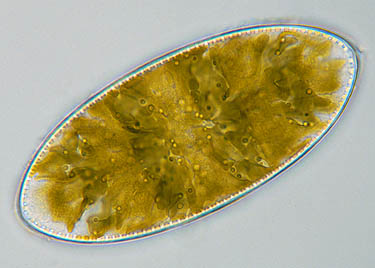
Yellow-brown chloroplasts can be seen in this Cymatopleura. |
|
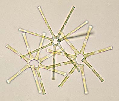
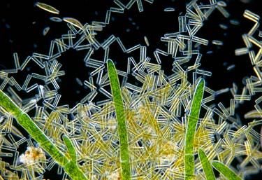 | Many species of diaom stay connected after the cells divide. They form colonies, long chains. Sometimes only the tips are connected and they form a zig zag pattern. |
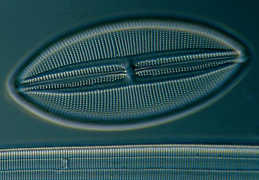
The cells of diatoms are ideal subjects for study under the microscope. They show complex patterns of very fine punctures and they often have all kinds of ornaments. The best way to make the beautiful structures of the frustule visible is to remove the content from the cell. Hydrogen peroxide can be used for this. But it is also possible to find empty frustules of dead diatoms. The extremely fine pores in the frustule of certain species of diatoms are used to test the resolving power of a microscope's lens. |
|
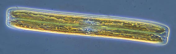
Diatom's locomotion Pennate diatoms, which developed later in the earth's history, are able to locomote in a slow gliding fashion in the direction of the length of the cell. The mechanism for this is still not well understood but it seems that through the slit alongside the cell (the raphe) tiny microfibrils protrude. With these they can move over a surface. In the picture of the cleaned diatom above the raphe can be seen as the thin central horizontal line. |
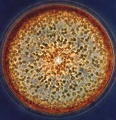
There are two different groups of diatoms, the pennates which are pen-shaped and the centric which are like a cylinder. In fresh water most diatoms you will see are of the pennate type. In marine waters the variety of body shapes is much greater. In the ocean they form the main part of phytoplankton, the photosynthetic organisms that float with the current. This image shows the top view of a large Coscinodiscus, a marine species that can just be seen with the naked eye. Clearly visible is the fine net-like structure of the siliceous cell wall. The yellow-brown chloroplasts used for photosynthesis are also easy to see. |
 |
| |
|
|
|
|
| Read the following MICSCAPE articles to find out more about a variety of diatoms (mainly marine) |
| Marine
diatoms 'Those who live in glass houses' |
use the POND LIFE IDENTIFICATION KIT !
or visit Wim's HOME PAGE
Microscopy
UK Front Page
Micscape
Magazine
Article
Library
all material © Wim van Egmond