|
Exploring classic insect test objects for the optical microscope: A subject which remains of interest to the amateur microscopist and to scientists in the field of photonics. by David Walker, UK |
Series Introduction: Using certain subjects as tests for microscope optics has a long history and had a vital role in the optical development of the achromatic compound microscope in the early 19th century. Some species of diatoms are well documented as tests and still very popular with enthusiasts, but using other subjects including animal hairs and various insect parts are also long established.
A selection of test objects in the author's slide collection will be shared in this occasional series in hope they are of interest. As others have noted, given the potential variability of a subject between mounts and the sometimes vague specification of the species, it's arguable if insect parts can be regarded as reliable test objects. But it's both great fun and instructive to inspect these slides while consulting the old books and to repeat the studies they recommended. The enthusiast nowadays can also try techniques developed since the Victorians such as phase and interference contrast if available.
| "The battle of the scales was fought out very fiercely about a dozen years ago, when Dr. Royston-Pigott claimed to have discovered beading in Podura and Lepisma scales and, although very little is heard of the matter now, it is not because it was settled, but because the combatants ceased from sheer exhaustion." T F Smith in "On the structure of butterfly and moth scales". Journal of the Quekett Microscopical Club, 1887, vol. 9, 178. |
Introduction
The distinctive silverfish was regularly seen in my youth in the 1950s and 60s, particularly in the bathroom where it often scuttled across the floor in a small flash of silver if disturbed. It prefers cool, damp, darkish habitats (see e.g. Wikipedia entry) but with now widespread central heating, the drier typical house may no longer favour this insect. 'Good riddance' some may remark as it is regarded as a pest because of its predilection for munching polysaccharides in book bindings, carpets, clothes etc. Although amateur naturalists like myself may likely eschew the 'find it and zap it' trend for many 'bugs' as we're always seeking something interesting to study. I haven't seen one for many years neither have British colleagues that I've asked.
It is an insect with a long history of study and was hoping to explore a live insect and in particular its scales both in situ and freshly removed but until recently have had to rely on a selection of prepared slides. The fine structure of its scales was used as an early test object from the 1820s when achromatic microscope optics were being developed but it remains a fascinating subject for the hobbyist to study. Insects such as the silverfish are being studied today by scientists using scanning electron microscopy and spectrophotometry to understand the nature of the structural silver effect and how such structures can be mimicked for various applications—the field of photonics.
Prepared slides
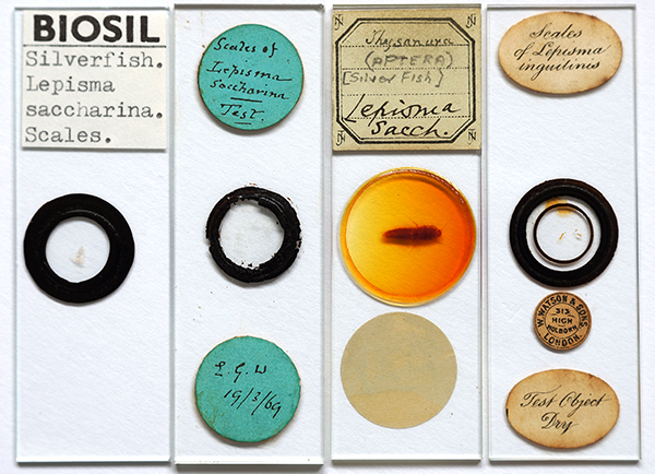
Prepared slides of the silverfish, often labelled as a test object can be found in many collections of old microscope slides. The slides I own which include a related species are illustrated below.
Above from left to right:
- Biosil slide ca. 1960s - 70s by the late John Wells. This is a dry mount typical of prepared slides of insect scales.
- A slide by 'L.G.W' dated 1869. Also a dry mount, the thicker glass slide makes it less suited for study with more demanding objectives / techniques.
- Whole mount. The 'JTN' in the label corners suggests it was made by J T Norman with two other hands adding further information. There is extensive oxidation of the balsam mount.
- A Watson slide (from the label format it was made between 1882-1908). It is labelled as a 'Test Object Dry' but have yet to find a reference specifically discussing 'Lepisma inguilinus' scales in this role. I'm not entirely certain what the modern equivalent species name is. If it is the same as 'Lepismodes inquilinus Newman' this is Thermobia domestica - the firebrat. A Mr. E. Newman reported 'A New Insect Pest in London in the 1863 'Popular Science Review'.
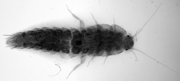
Above. The whole mount of Lepisma saccharina taken under a stereo (total optical mag ca. 12X). A heavy red cast in the mount had to be digitally removed hence the uneven lighting. This type of slide is useful for e.g. studying fine structure of appendages but in this case provides little useful information on the scale layout nor on the insect's 3D structure compared to studying an unmounted insect. The structural silver colour is of course lost.
Above. Scales of Lepisma inguilinus on the Watson slide shown. Now believed to be Thermobia domestica, the firebrat. Brightfield Zeiss 25/0.60 objective with a little off-axis oblique. Post capture tonal balance increased for these intrinsically 'flat contrast' subjects.
Historical summary of Lepisma studies using the microscope
A timeline of selected Lepisma studies under the microscope and other key dates are summarised below.
| Date | Observer | Comments | Reference (most accessible on archive.org) |
| 1665 | Robert Hooke | Observation LII, large detailed engraving of whole insect, scale discussion but not on fine structure. Were his compound microscopes good enough to resolve scale fine structure? Hooke makes perceptive comments about the nature of the scale reflectance and proved to be 'essentially correct' by modern EM studies, see 2001 entry. |
Micrographia |
| e.g. 1702 | Anthony van Leeuwenhoek | No record found of Lepisma study but he described and illustrated fine structure on insect scales e.g. the wing scale hairs of the cultivated silkworm Bombyx mori. See Micscape June 2012 article. |
The Collected Letters of Antoni van Leeuwenhoek. Vol. XIV, 1996. Letter no. 236 [146], 20 April 1702, pp.101-133 and plates VII and VIII. |
| 1758 | Linnaeus | Assigned the silverfish the scientific name Lepisma saccharina in the Order Aptera. | Systema Naturae 10th edition |
| 1825-1827+ | Dr. C R. Goring | Insect scales are reported as one a number of types of useful test objects for the achromatic objectives being developed. Notably pioneered by Dr. Goring. Quekett (1848, see below) stated that Goring was prompted to 'adopt' them as tests after reading Van Leewenhoek's silk moth wing scale studies (see above). | One of his major papers is in The Quarterly Journal of Science, Literature, and the Arts, 1827, Vol. XXII, pp.265-284. Plus Plate. |
| 1828 | G. Dakin | Earliest paper found to date that specifically mentions Lepisma scales as a test object. Uses incident light. "Dr. Goring uses the scales of the Lepidoptera as test-objects: but the most beautiful and delicate test-objects that I have ever seen are the scales of the Lepisma saccharina: they are so very thin, and the lines on them so very fine, that they will bear almost any power." (Page 431 of ref.) |
The Philosophical Magazine or Annals, 1828, July December, vol. IV, 429 - 431. |
| 1829 | Thomas Gill (ed.) | Earliest paper found to date that includes both an illustration of the scale and a description. Notes the diverging lines on the scales. Specimen supplied by Mr. T. Carpenter who was credited regularly in the early papers for supplying other classic test scales, e.g. of the blue butterfly Morpho menelaus and springtails ('Podura'). Links to March 2015 and Feb. 2011 articles in Micscape respectively. | Gill's Technological Repository,1829, vol. IV, p.72 and plate II, fig. 26. (Scale obscured by folded plate on archive.org.) |
| 1832 | Andrew Pritchard | Chapter XVI devoted to an extensive discussion of 'Test Objects' including insect scales and mammal hairs. Classifies them into three 'Sections' - 'Easy, Standard, Difficult' according to Penetration which he associates with 'angle of aperture', i.e. NA and resolution. Lepisma is classed as 'Easy'. | The Microscopic Cabinet, Chapter XVI, pp.135 - 161. Plate 12 devoted to tests, Lepisma fig.1. |
| 1848 | John Quekett |
Chapter XX devoted to an extensive discussion of 'Test Objects' plus Plates 7,8,9. Extends Pritchard's studies. Notes that the improvements in achromatic objective are reflected in the superior illustrations and detail. Describes the finer structures of some diatom frustules and manmade grating plates as becoming the more demanding tests. |
Practical Treatise on the Use of the Microscope, pp.422-447. |
| Various papers in the following period, some were dubbed 'the battle of the scales'* discussing the apparent fine structure of the scales e.g. the 'beading' seen by some workers and whether real or artefacts. Only a selection shown. Generally agreed that the coarse 'beading' was an artefact as there can be parallel coarse ribs along the long axis on one side and radiating ribs on the others, which can 'interfere' in transmitted light. Although other papers discuss the possible fine structure on the membranes between the ribs. |
*See quote at the head of article by T F Smith in 1887. | ||
| 1865 | Richard Beck | Includes a superb plate illustrating fine structure (see below). Uses Lepisma scales as an example that transparent objects with structures both sides can give artefacts in transmitted light. Describes scale removal from insect. How water introduced under slip affects apparent structure. | The Achromatic Microscope, pp.49-53, plate XIX devoted to multiple aspects. |
| 1873 | G. W. Royston-Pigott | One of the main proponents of the 'beading' seen. In this paper, the coarse beading by rib crossing from either side of scale interfering are dismissed but reports much finer beading between the ribs. |
On the Spherules which compose the Ribs of the Scales of the Red Admiral Butterfly (Vanessa Atalanta) and the Lepisma Saccharina, 1873, The Monthly Microscopical Journal, Vol. IX, 59-65, Plate XI. |
| 1874 | John Anthony |
Reflected light studies supports beading as artefact. Reports fine structure between ribs but I am uncertain if genuine. Published SEMs seen are not at a high enough mag to confirm. |
The Scales of Lepisma as seen with Reflected and Transmitted Light. The Monthly Microscopical Journal, 1874, Vol. XI, May 1st, 193-195 and Plate LIX. |
| 1874 | G. W. Morehouse | Careful study with observations of the coarse structures through to possible fine structure between ribs. Notes 'transverse corrugations of the membrane". | The Structure of the Scales of Lepisma Saccharina, The Monthly Microscopical Journal, Transactions of the Royal Microscopical Society, 1874, vol. XI, 13-15. |
| Later 19th and 20th century | In amateur circles interest in Lepisma scales as a test subject decreased as no longer regarded as a demanding test object. | ||
| 2001 | M. C. J. Large et al | A modern EM study (a silverfish in the Ctenolepisma genus) of the scale structure in the field of photonics where the physical structures and their interaction in light are now of great interest. | M. C. J Large et al, The mechanism of light reflectance in silverfish, Proc. Roy. Soc. A, 2001, Vol. 457, pp. 511-518. Kindly offered by the authors for public access as a pdf on researchgate.net. |
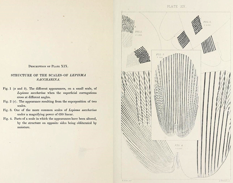
Richard Beck's superb plate from his 'The Achromatic microscope' pub. 1832 where he notes that the 'interrupted appearance (described as 'beading' by some workers) seen is an artefact. In the public domain at www.archive.org
The insect featured as Observation LII 'Of the small Silver colour'd Book-worm' in Robert Hooke's classic illustrated text 'Micrographia'. The splendid engraving from 'Schem: XXXIII' is shown below. Its scientific name Lepisma saccharina in the order Aptera was assigned by Linneaus in the 10th edition of his 'Systema Naturae' 1758.
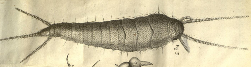
Above: 'Schem: XXXIII Fig. 3' - the 'small Silver color'd Book-worm' illustrated in Robert Hooke's 'Micrographia' 1665. Source: From a copy on www.archive.org in the public domain.
Live specimen
I inquired on social media about other people's experiences of finding silverfish and a UK microscopist found one at work and kindly sent the live specimen (named Sylvester) in the mail in a vial with damp cotton wool. It survived the trip and has been living in a Petri dish in the dark with damp paper for food. On receipt it looked a young pale specimen but has grown over a year to a darker brown colour. The ones I vaguely recall from my youth were a marked silver colour although Google images do tend to show many as a darker brown; possibly variations between sexes, comments welcomed. It was photographed quickly under the bright lights with a stereo before returning to its home. Its scuttling around the dish suggested that this nocturnal animal did not appreciate fame in the bright lights. This example has a body length of ca. 8.5 mm.
Collecting scales from a live specimen: I didn't want to kill this obliging specimen for scale studies so initially relied on the prepared slides owned. I later found that if the silverfish was allowed to scurry along with a coverslip lowered so that it gently touched its back, a number of scales were released. This allowed a scale strew with a known orientation to be collected; scales top surface uppermost if coverslip used and undersides uppermost if a slide was used instead to touch its back. A clean slip then covered the scales for the latter.
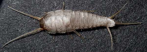
Live silverfish, body length ca 8.5 mm. Olympus TG-5 in Microscope mode, single image. Attempts to use its internal focus stacking mode was unsuccessful as would not stay still.
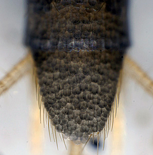
A higher mag of the tail on a stereo shows the overlapping scales.
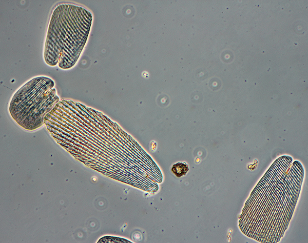
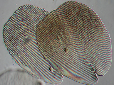
Scales from live specimen scales uppermost, as on body, dry mount. Zeiss 16/0.40 Neofluar phase. Horizontal field width (HFW) ca. 395 microns.
The variety of shapes, sizes and rib coarseness can be seen. (Also see Biosil prepared slide below.)
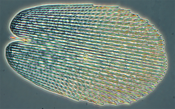
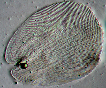
Scale from live specimen scales uppermost, as on body, dry mount. Zeiss 40/0.70 Neofluar phase. HFW ca. 140 microns.
The beading artefact can be seen as radiating ribs on the underside interact with the parallel ribs uppermost.
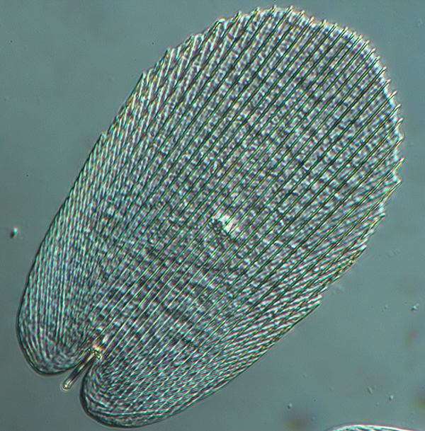
As above but DIC. The apparent scale structure changes as the scale is rotated with respect to the DIC shear axis (NW-SE in this case).
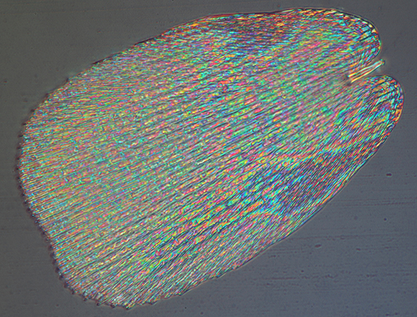
Scale from live specimen with underside of scale uppermost, dry mount, uncovered. Zeiss 40/0.85 epi DIC. Two image stack to show the pedicle (insertion shaft of the scale top right) and the plane of the scale). The colours may be interference between the upper and lower scale layers?
Modern electron microscopy studies and photonics
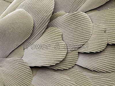 Our forbears didn't have the benefit of electron microscopy to answer any uncertainties of microstructure for subjects viewed under the optical microscope. The SEM image right is ©Power and Syred, courtesy
of Andrew Syred at PSMicrographs, which shows the overlapping Lepisma scales in situ on the insect. The radiating rib structure is shown on the upper surface with no evidence of beading. Other examples of SEM imagery of the scales and insect can be found e.g. at the Science Photo Library.
Our forbears didn't have the benefit of electron microscopy to answer any uncertainties of microstructure for subjects viewed under the optical microscope. The SEM image right is ©Power and Syred, courtesy
of Andrew Syred at PSMicrographs, which shows the overlapping Lepisma scales in situ on the insect. The radiating rib structure is shown on the upper surface with no evidence of beading. Other examples of SEM imagery of the scales and insect can be found e.g. at the Science Photo Library.
The fine structure of insect scales attracts a lot of attention in the field of photonics. Large et al (see ref. dated 2001 in table earlier) presented fascinating studies including SEM and TEM of the nature of the broad band reflectance seen in silverfish. They showed that reflectance in the visible was mainly caused by multilayered structure in the upper layers of exoskeleton with the scales only modifying some parts of the spectrum. Noting that scale loss was believed to be a defence mechanism it would be beneficial to the insect if such losses did not cause colour changes to make it more conspicuous to predators.
Could Hooke have observed the Lepisma scale fine structure?
Large et al also noted that Robert Hooke (see Timeline) made the first studies and that he makes perceptive comments about the nature of the scale reflectance and which has proved to be 'essentially correct' by modern EM studies. Hooke's superb engraving in Micrographia does not seem to show evidence of scale fine structure (other than as part of the engraving method). But it is interesting to speculate if he could have potentially resolved the fine structure with his compound microscope. The objective NA required to resolve the coarser ribs is modest ca. NA 0.06 and have previously shown in a Micscape article (January 2015 and image from article below) how such scales can be useful for assessing the resolution of older optics such as the typical set of single lenses provided e.g. in Cary-Gould type microscopes.
To date, I have been unable to find reports of the resolution of extant optics in compound microscopes of Hooke's period (the lenses were often missing and replaced later), but Van Cittert in his seminal study (1) reports a typical resolution of 10 microns for the four objectives giving a total mag of 28 - 100X in a later Culpeper compound microscope. The historians note that there was little development of microscope optics for compound microscopes in the short term. The coarsest scales are just under 5 microns apart so this suggests that Hooke's compound microscope(s) if comparable could not have resolved the fine structure.
Brian J Ford, a leading historian of microscopes and user of period models has proposed (2) that Hooke must have used single lens microscopes to record the fine details shown in some subjects illustrated in Micrographia, his compound microscopes being unable to resolve such structures. If Hooke was able to make single lenses comparable to the more modest lenses made by Van Leeuwenhoek which Van Zuylen studied (3), he found that a Van Leeuwenhoek 74X lens had an NA of 0.13. Its measured resolution was 4 microns with a 110X lens resolving 2.3 microns. So Hooke may have seen Lepisma scale fine structure if such lenses were used (more likely if scales were removed for transmitted studies than in situ on the insect?). If he did observe such structure he made no report of it in the text or suggested in the superb illustration. This is all speculative of course, comments welcomed!
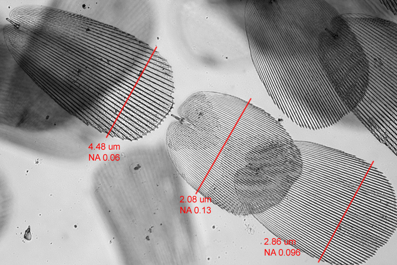
Lepisma scales, Biosil slide, Zeiss Neofluar 16X/0.35 brightfield (from the June 2015 article, 'An 1830s Cary-Gould style microscope by Carpenter and Westley: Exploring the versatility and optical performance of a popular single lens / compound microscope'). The figures are for the average rib separation measured along the red line on each scale and corresponding lens NA required to resolve.
The author David Walker is an amateur microscopy enthusiast not a historian and welcomes any comments / corrections.
Acknowledgements: Many thanks to the British microscopist who kindly supplied the live specimen of silverfish which continues to thrive. Thank you to Andrew Syred at PSMicrographs for permission to share the splendid SEM image.
Additional References not in Timeline: Many of the older journals and books are available on www.archive.org.
1) P. H. van Cittert, Descriptive Catalogue of the Collection of Microscopes in charge of the Utrecht University Museum with an introductory Historical Survey of the Resolving power of the Microscope, 1934 P. Noordhoff N.V., Holland. Chapter III, Chromatic compound Microscopes, G. The Culpeper microscope (dated 'first half of the 18th century), table, p.37. Objectives nos. 2-5, 28X - 100X, resolution, 1/100 mm i.e. 10 microns.
2) Brian J. Ford, Advances in Imaging and Electron Physics, 2009, Elsevier Inc, Chapter 2, Did Physics Matter to the Pioneers of Microscopy?, section 6.2 Sources of Inspiration, p.77.
3) J. van Zuylen, 'The Microscopes of Antoni van Leeuwenhoek', J. of Microscopy, 1981, 121/3, pp. 309-328. (Reprinted in 'Antoni van Leeuwenhoek 1632-1723', Eds. L. C. Palm and H.A.M. Snelders, Rodopi, Amsterdam, 1982, pp.29-55.) Table 1 summarises the properties of nine remaining Leeuwenhoek microscopes.
Detailed and general resources on microscope test objects
C. R. Goring, 'On Mr. Tulley's thick Aplanatic Object Glasses, for Diverging Rays; with an Account of a few Microscopic Test Objects', Quarterly Journal of Science, Literature and the Arts, 1827, XXII, 265-284. (Link is to Google Books beginning of paper.)
C. R. Goring, 'On Achromatic Microscopes, with a description of certain Objects for trying their Defining and Penetrating Power', Quarterly Journal of Science, Literature and Art, 1827, Jan-Jun (XXIII), 410-434. (Link is to Google Books beginning of paper.)
S. Bradbury, 'The Evolution of the Microscope', Pergamon Press, 1967. This book provides a detailed but very accessible discussion; chapter 5, 'The Development of the Achromatic Microscope'.
G. L'E. Turner, 'The Microscope as a Technical Frontier in Science', in ''Historical Aspects of Microscopy, Eds. S. Bradbury and G. L'E. Turner, The Royal Microscopical Society, 1962, pp.175-199. Figs. 9-11 plot the increase of numerical aperture and resolution of test objects with year showing the particularly rapid improvement from 1830s to ca. 1860.
W. B. Carpenter, 'The Microscope and its Revelations', London, 1857, second edition. Section 108, 'Test-Objects', 183-189.
Cheese mites and other delicacies: the introduction of test objects into microscopy, Endeavour, Volume 27, Issue 3, September 2003, 134-138, Jutta Schickore. (Link is to an Abstract and full access to subscribers.)
J. W. Griffith and A. Henfrey, 'The Micrographic Dictionary', third edition, 1875, p.773 - 776, ('TEST-OBJECTS' entry). (Link is to www.archive.org full download of the 1856 first edition.
A. Pritchard, 'An Account of Test Objects for Microscopes', The London and Edinburgh Philosophical Magazine, 1833, 3rd series, vol. II, 335-342 and Plate III.
A. Pritchard and C. R. Goring, 'The Microscopic Cabinet ...', 1832, London.
S. Bradbury, 'Test Objects', Quekett Journal of Microscopy', 2002, 39, 215 - 224.
J. Schickore, 'Test objects for microscopes', History of Science, 2009, 117 - 145.
Published in the June 2020 edition of Micscape.
Please report any Web problems or offer general comments to the Micscape Editor .
Micscape is the on-line monthly magazine of the Microscopy UK web site at Microscopy-UK
© Onview.net
Ltd, Microscopy-UK, and all contributors 1995 onwards. All rights
reserved.
Main site is at www.microscopy-uk.org.uk
with full mirror at
www.microscopy-uk.net
.