One of the problem about having a fecund mind–I love that word, fecund, fecund, fecund, it sounds dirty, but it simply means “fertile-- is I have always concocted more projects than I could ever deal with; some get started, some are still in an embryonic state. I’m talking literally hundreds of projects here–FECUND! Another word I love which was a favorite of W.C. Fields is kumquat. It’s a deliciously eccentric word–kumquat, kumquat, kumquat. Well, enough of that for now.
My first series of projects developed when I was a teenager and they centered on protozoa and incidentally some algae. I got so focused on these fascinating organisms that I began to devise my own primitive taxonomic charts. From a scientific point of view they were quite useless; but, on a quotidian (another one of those wonderful eccentric words. It just means “daily”) basis, these primitive charts helped me recognize different groups of protists and understand some of their interconnections. More importantly, this process got me interested in formal taxonomy and I came to understand both its benefits and its absurdities. Taxonomy is anything but precise and is sometimes downright silly. However, its importance, when working properly is that it provides a set of common reference points for communication and the names are sometimes helpfully descriptive. Imagine the difficulties we could have if my friendly rival, Erik Klondiker, referred to a particular protozoan as Klondiker splendida and I used the name Magnificatus howeyii for the very same organism. Not only would that create confusion but, if we ever discovered that we were both referring to the same organism that might lead to a blood feud. As it turns out, synonymy is a significant aspect of the taxonomic enterprise. Because of the snail pace of communication prior to the mid-20th Century, consistency was often a difficult goal to achieve. Can you believe it, I remember writing letters by hand with a goose quill dipped in an inkwell—well, O.K., a bit of hyperbole, but I did use a fountain pen and put a stamp on an envelope, walked down to the end of the block and put it into a mailbox and then perhaps 6 weeks later, I would get my Arf and Nonnie Cosmic Secret Decoder Ring.
Now, with the advent of the Internet and digital cameras, I can take an image and send it off to a friend or an expert or an expert friend and may possibly get a response within minutes. This is assuming that he and his wife or she and her husband or he and his husband or she and her wife are not otherwise occupied away from the leash of their computers. So, now we can have real-time disputes about the intricacies of taxonomy. For my part, I say, “No thanks! Recent taxonomic developments have gone to such extremes that I have quite lost interest in the enterprise as it is conceived of at present.
My next teenage obsession was, oddly enough, tunicates, although I lived 2,000 miles from any ocean, over 1,000 miles from any gulf or sea so, an odd sort of fixation. A college textbook on invertebrate zoology was the culprit. I happened on a chapter on Urochordates. Although much of it was beyond the grasp of my teenage brain, I was utterly fascinated by the catalog of oddities associated with the tunicates, perhaps because I felt myself to be such an oddity.
The next great onslaught of projects came from a summer visit to the coast of Maine. Some dear friends invited me to spend a week to two with them at their house which was only a stone’s throw from the ocean. What self-respecting amateur naturalist could resist such an invitation? The night before I was to leave for Maine, they called to tell me that husband was ill and had to have surgery, but asked if I would go anyway and act as caretaker. There had been vandalism in the area and it would only be a matter of a month. A month of uninhibited access to a beautiful coastline with tidepools, shallow coastal inlets, and mudflats ripe with burrowing worms and mollusks. I pack a stereo microscope, bottles, nets, preservatives and anesthetizing fluids and left the next morning. What a marvelous summer, collecting each day and discovering wonderful creatures that I had previously only read about and seen drawings of. Needless to say, I returned with many preserved specimens and plankton samples. For a fuller account of that summer, you might look at my earlier article.
I suppose the next great obsessive project was the discovery of Lacrymaria olor and the attempts to culture it. Here is a creature that has an extensile neck that it can extend up to 10 times its body length!–very handy in crowds. My friend Mike and I went through combination after combination of culture media in the effort to find a medium that was satisfactory. We even went to the extent of including powdered milk in some recipes which after sitting for a bit has the revolting reek of baby vomit. The final formula which we adopted provided reasonably stable cultures for several years which allowed for some unorthodox and interesting experiments, one of which involved the use of a mutagen and produced 3 headed specimens!
About this time, I acquired a splendid Wild-Heerbrugg inverted phase microscope. I also acquired some algae and detritus samples from aquaria in a local pet shop. One afternoon, I was looking at a sample of it and saw a bit of pinkish-red detritus and then noticed that there were such bits all over the bottom of the dish and my first thought that it was just some sort of odd nuisance until I saw one slowly move! I increased the magnification on my stereo-zoom and began taking a series of pictures which revealed cilia distributed over the surface. I was fairly certain that it was not some sort of weird amoeboid form, partly because it appeared to be multi-cellular. I took my images up to the Zoology department at the university along with a sample of the organism. I recruited 3 of my colleagues to give me their opinions regarding this wee, weird beastie. One colleague was a protozoologist and cell biologist, the second was trained as a marine biologist, the third was a geneticist. Surprisingly, I got 3 radically different opinions. One thought it might be a bryozoan, another that it was the larva of a flatworm, and the third that it was a developmental stage of an unusual sponge. Each made a convincing argument for his position, but wet-behind-the-ears, arrogant, upstart that I was, I came away unconvinced. I went home and spent hours and hours looking at this strange thing. It’s small–usually about 1 to 3 mm and is quite flat, varies in color from a light gray to deep pink. It didn’t seem to have much of anything in the way of differentiated structure beyond the fact that it was indeed multi-cellular. I began searching the literature again. Previously, I had looked at sites which were quite technical, but this time, I decided on a different approach. I started looking though textbooks on invertebrates. After several unsuccessful attempts, I came across a brief chapter in Barnes’ book on invertebrate zoology. And there it was–Trichoplax adhaerens, the only member of the phylum Placozoa. How much stranger than that can you get than to require your own phylum? Clearly, another extra-terrestrial critter–probably from Titan, the largest moon of Jupiter. So, definitive proof that not all weird extraterrestrials are green. My enthusiastic identification was not shared by my colleagues. In general, academics hate to be wrong about even the smallest matter (and, remember, Trichoplax is only 1 to 3 mm in size). Interestingly, many of the collections of Trichoplax were in marine aquaria. I wrote a little article on this strange, wee critter which appeared in Micscape.
Shortly after it appeared, I began to get emails and phone calls from researchers wanting samples to start their own cultures several of whom wanted to do gene sequencing. Unfortunately, I had by then moved on to other projects and no longer had any specimens available.
Next came a period of obsession with echinoderms which has never really dissipated and reappears vigorously from time to time. This is a phylum that demonstrates the power and the importance of classic and moderate modern taxonomy. One can immediately understand grouping the ophiuroids (brittle stars) and asteroids (sea stars) together. But, incorporating the other three major groups seems a real stretch. What would lead any rational soul to claim sea cucumbers, sea urchins, and crinoids belong together with starfish and brittles stars? Preposterous! Sea cucumbers–slimy bags full of sand from which they extract tiny bits of food. Sea urchins–spiky, living pincushions which have a hard test (shell) beneath those prickly spines. Crinoids–the multiple-armed elegant sea lilies and basket stars. Only an idiot would want to deposit all of these in the same phylum–actually it’s a whole bunch of very bright idiots who accept this taxonomic arrangement. But why? Much of the answer lies in embryology and morphology.
The larval stages of these 5 groups demonstrate special developmental characteristics which are highly convincing and which you can discover for yourself with some good library and Internet research.
The key morphological aspect turns around pentagonal symmetry (generously interpreted with allowances for significant exceptions). The classic model for starfish and brittle stars is five arms radiating from a central disk.
As it turns out, although it’s much more difficult to determine immediately, pentagonal symmetry is a crucial aspect of the other 3 groups as well. If you start poking around a sea cucumber, you’ll quickly discover 5 double rows of tube feet and 5 groups of oral tentacles. Naturally, Mother Nature has created a few exceptions to keep us on our toes and thereby admonishes us to pay attention to detail.
Next, take a look at one of those spiny sea urchins and you may well wonder how pentagonal symmetry can be any part of such a creature. Well, pick out a nice specimen and prepare yourself for the thrill of vandalism. The first step is to strip all the spines off of the test. Ouch! That was me, not the sea urchin. And, what do we find? What we often find is a structure that is both beautiful and amazingly complex. Also we find 5 pairs of double rows of holes through which the tube feet extend when the organism is alive. Also, there are 5 paired rows of ambulacral plates and 5 pairs of interabulacral plates as well. So, we’re doing very well in terms of pentagonal symmetry. There is also the structure of the mouth, a complex mechanism composed of calcareous plates. There are 5 teeth which are used for scraping algae off the rocks or substrate. The whole structure is known as Aristotle’s lantern since he was the first to discover and describe it. In addition, there sets of a number of different kinds of plates (5 each) which support the muscles that control the movement of the teeth.
With crinoids, it is also relatively easy by looking carefully to discover pentagonal symmetry in spite of all the elaborate branching.
So, from these examples, we learn that it is not only crucial to observe meticulously, but that we also need to sharpen our ability to see the elaborate connections between apparently disparate organisms and realize that they may be closely related. To do this, one could devote a life time to the study of any one of these groups. Many, many unfinished projects which I have involve these amazing creatures and I hope to pass on some of these projects to former students.
My next extended obsessive period is one which also keeps reasserting itself–crystals under polarized light. A good starting point for this project is Ascorbic acid (Vitamin C). This is a very versatile substance that produces a wonderful range of results and has the great virtue of being easily combined with other chemicals to produce some spectacular and often unexpected results. Make up solutions in various concentrations, place a couple of drops on a slide and keep track of the concentrations and results. Temperature will also produce some interesting changes in the patterns of crystallization. If you don’t have a warming table, then dissolved the Ascorbic acid in tap water at various temperatures again keeping careful records. You can also put a small try of such slides in a freezer compartment of our refrigerator for different lengths of time.
Then, even more fun ensues, when you start mixing with some other chemicals. However, a word of caution, be certain that the things you mix will not produce some nasty reaction. So, do indeed experiment, but do so rationally and carefully. Now, I will indulge myself and give you series of micrographs of Vitamin C, some other reagents, and some combinations with only the briefest notes.
WARNING! Once you start these explorations, there is a high probability of addiction and there is no antidote short of selling your microscopes! I will concretely demonstrate that fact for you by here including 437 images carefully selected from several thousand. (Just kidding. But I will give you 30 images and, yes, I do have several thousand.)
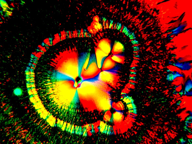
This is a combination of Ascorbic Acid and the biological stain, Methylene Blue. Over the years, I have found that a variety of stains can when mixed with other reagents producing some quite striking results with polarized light.
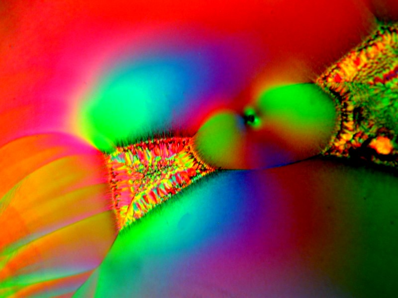
This is another example where this time the combination is Ascorbic Acid and the biological stain Light Green.
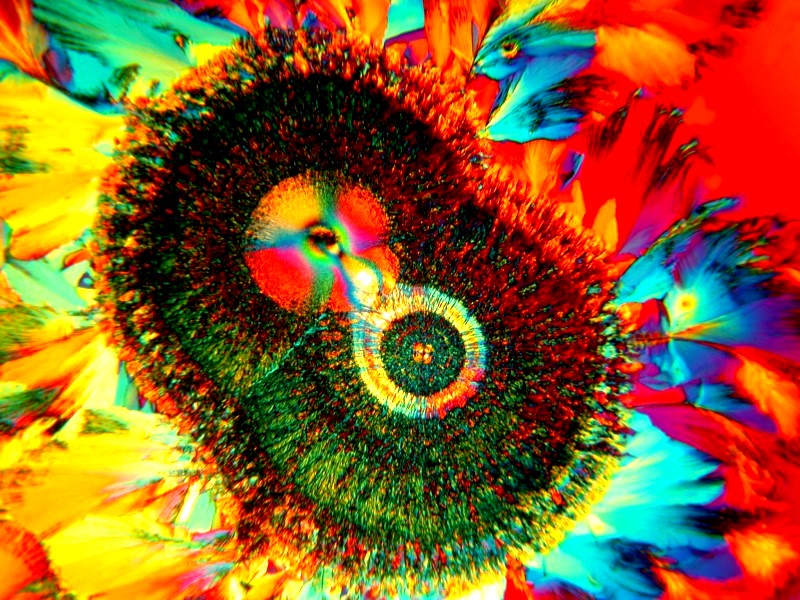
Another Ascorbic and Methylene Blue
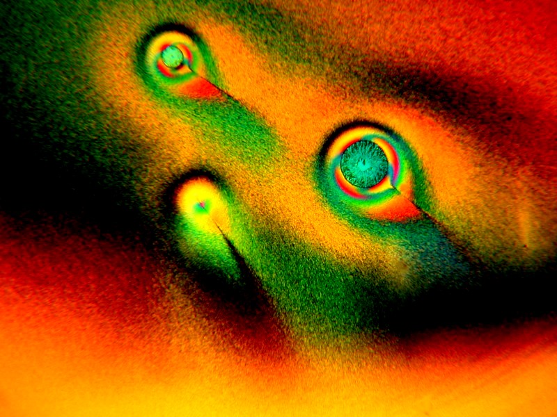
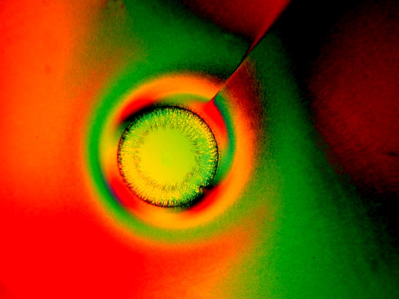
The 2 images above are a combination of Ascorbic and the biological stain Toluidine Blue. Once again, I urge you to try dilute solutions of stains as they often produce interesting results. You might also try various sort of inks.
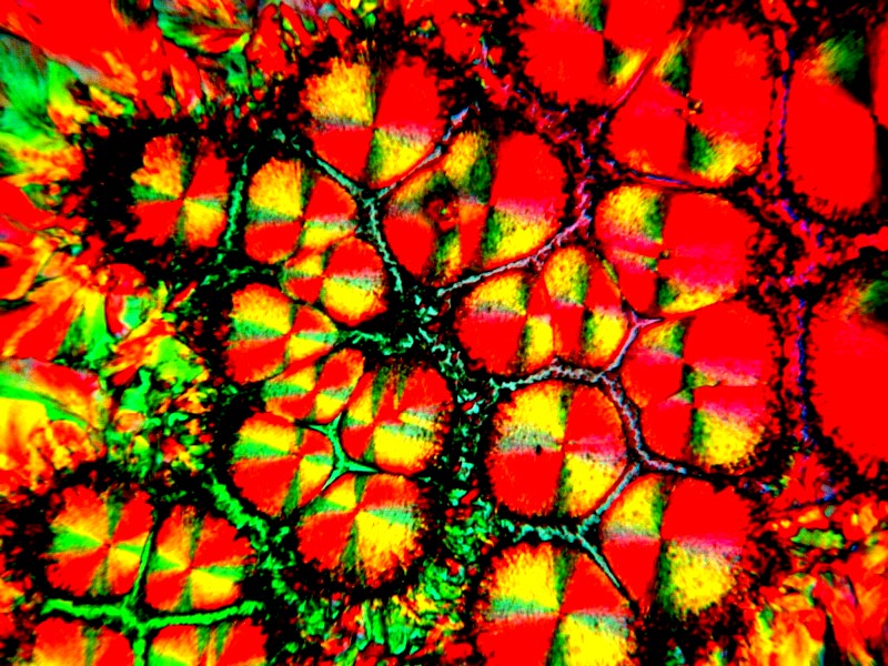
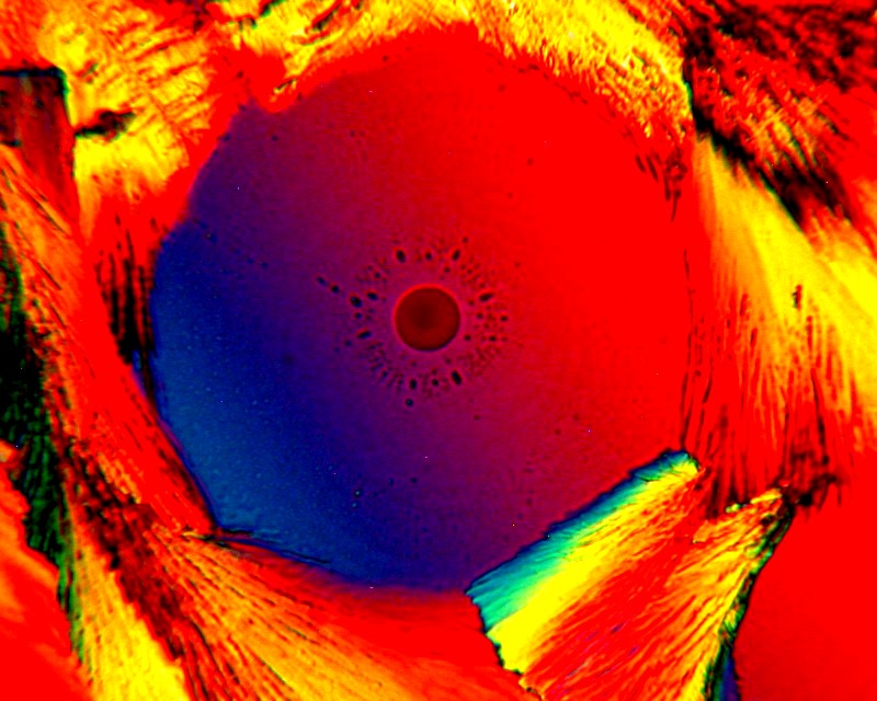
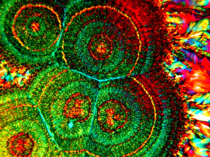
These 3 images again employ a stain with Ascorbic, this time an intriguing combined stain called Indigo Carmine.
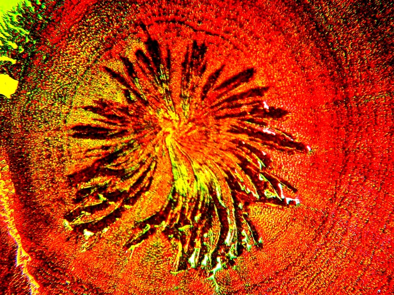
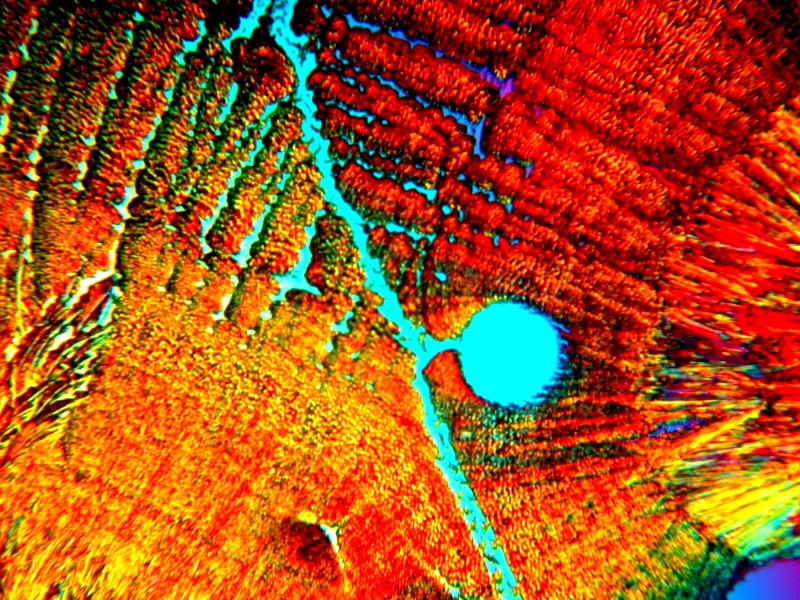
Sometimes a common substance which is easily obtainable or can be found around the house can surprise us. These 2 are Ascorbic mixed with Citric Acid.
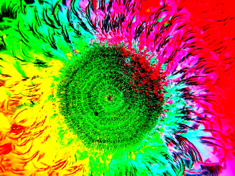
This is a very easy example of what can turn into a whole series of fascinating images. Here I simply added a drop of 70% isopropyl (rubbing) alcohol to a drop of Ascorbic. The alcohol and the water interact and the whirling creates interesting patterns. This is worth trying with other mixtures such as those employing stains.
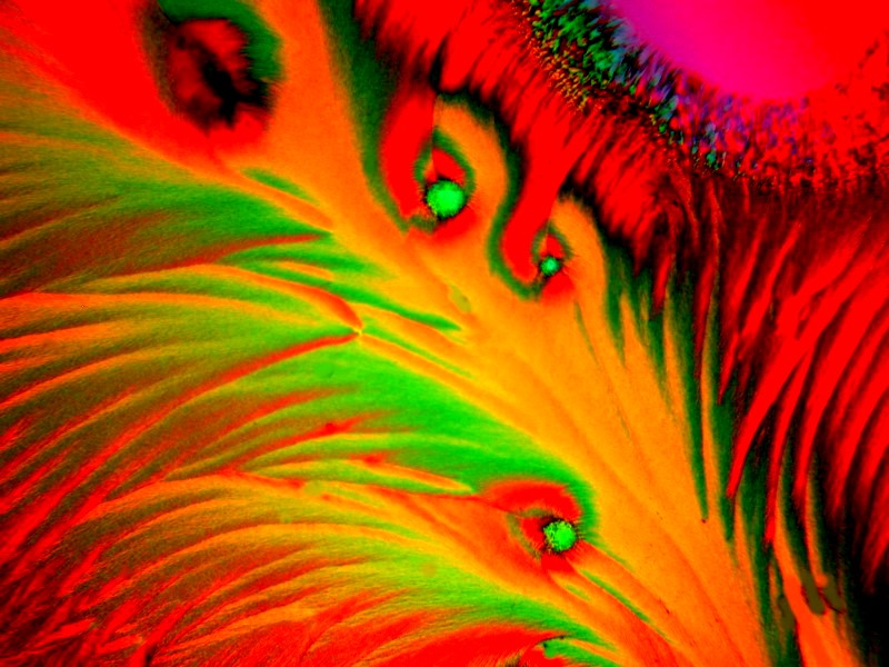
This is a more complex mixture using Ascorbic, Prozac, and the stain Neutral Red.
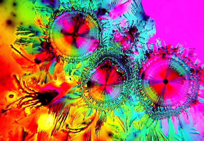
Ascorbic plus Crystal Violet. Crystal Violet is never a completely “pure” dye and the small residues of other substance in it make it versatile, unpredictable, and thus great fun to try.
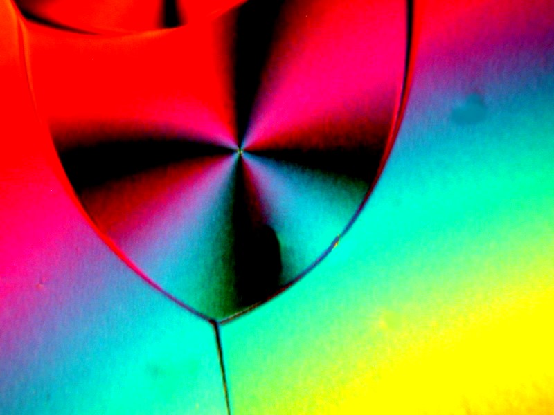
This is a mystery image. It is Ascorbic Acid mixed with some other substance which remains mysterious, because I can’t read my handwriting.
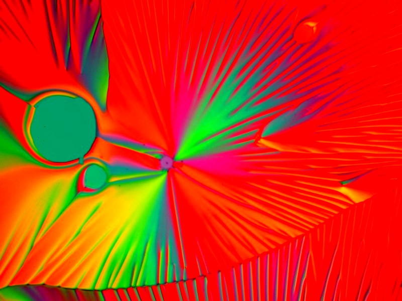
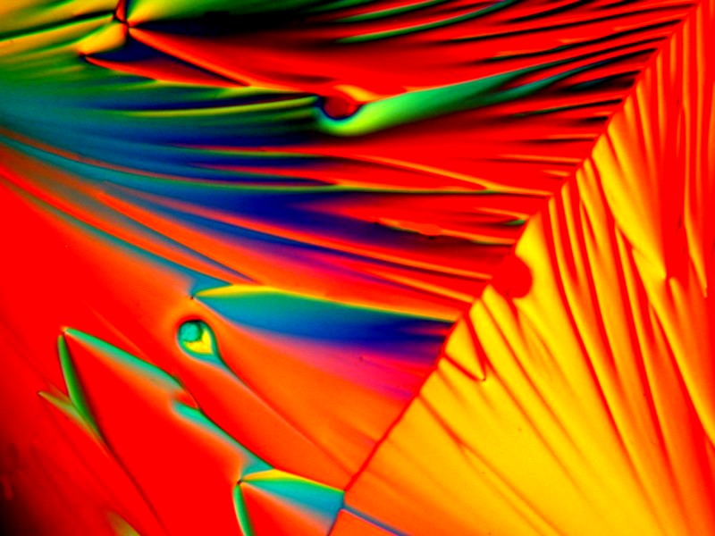
When you get to my age, it is necessary to soak ones feet from time to time and an effective way to do that Is to add some Epsom salts (Magnesium sulfate) to warm water. Some substances typically produce rather thin, delicate needle-like crystals, whereas Magnesium sulfate tends to produce those, but rather geometric forms as well. These 2 are both Ascorbic and Magnesium sulfate.
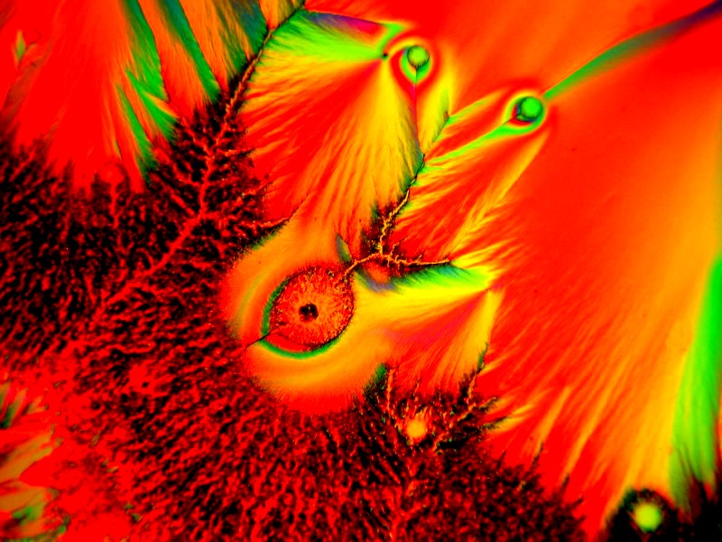
This is a lonely image with no friends to mix with; it is simply Ascorbic Acid.
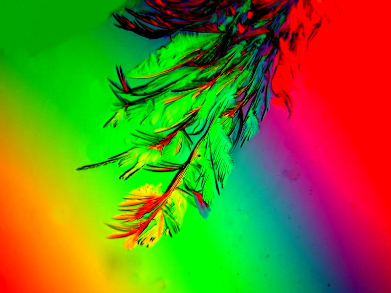
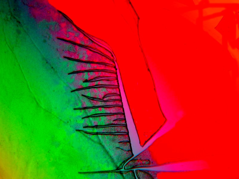
Two images with Ascorbic mixed with both Vanillin and Methylene Blue.
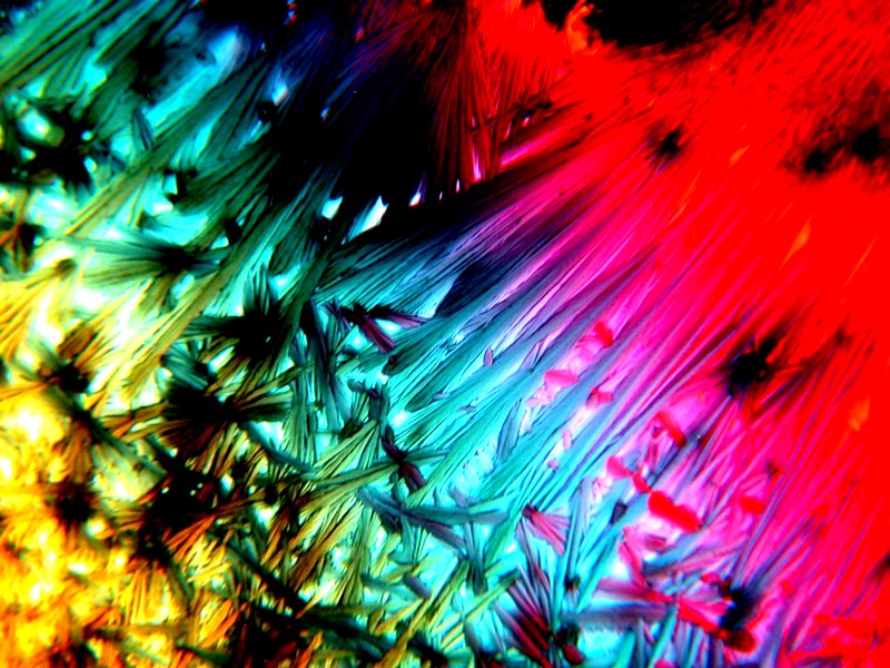
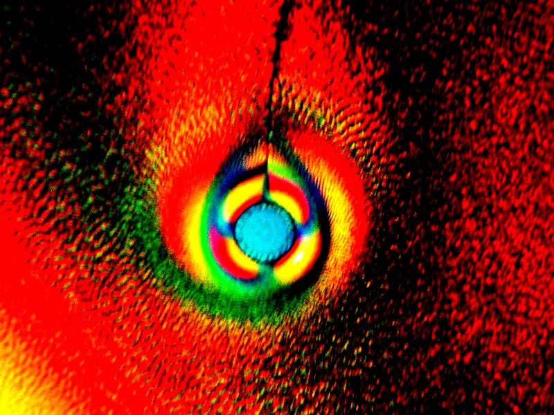
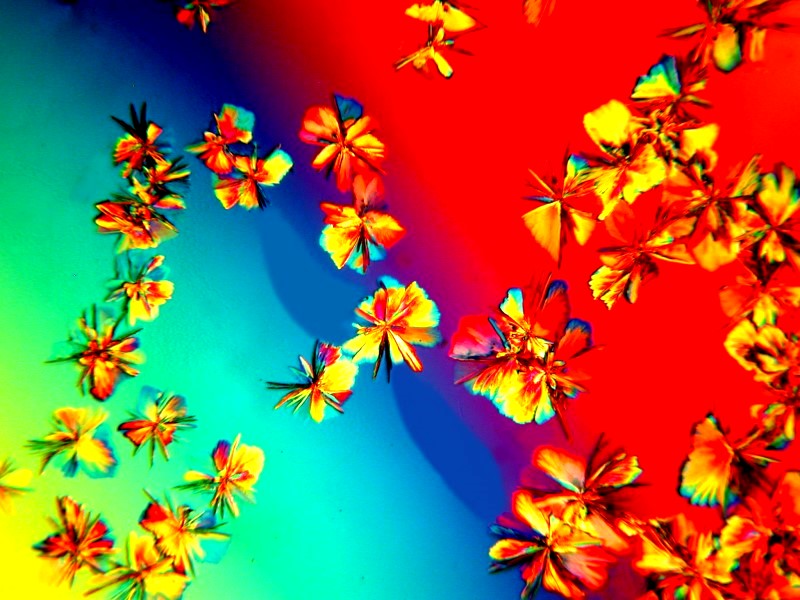
There is a prescription drug called Metformin which is used for diabetes type 2. I had a bad reaction to it had some left over which immediately went into my lab for crystal experiments and here are 3 results above which are Ascorbic and Metformin. However, I will be presenting you with some other images using Metformin without Ascorbic in a minute.
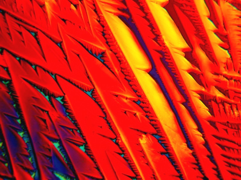
Here is another one of my experiments with isopropyl alcohol, this time mixed with Metformin.
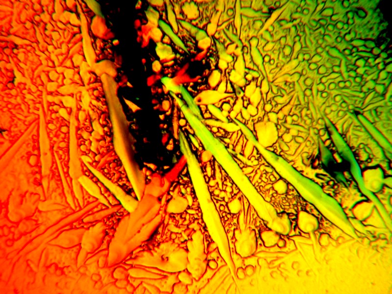
Campden is a tablet, bottles of which you can buy for making wine and brewing beer. It is sodium metabisulfite.
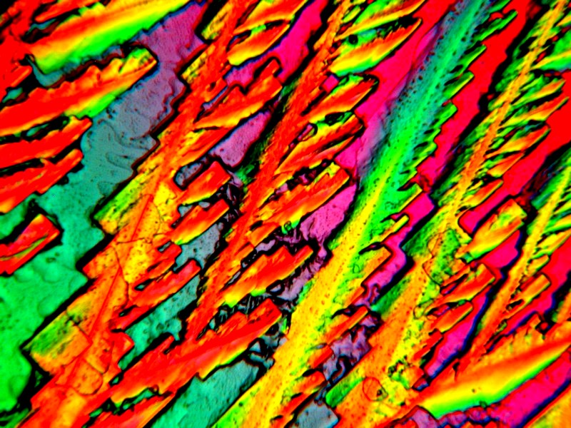
Metformin combined with a drop of a nose drops (no, not from my nose, from the little bottle of drops to clear the congestion in my nose).
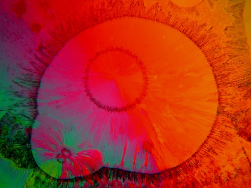
Blood pressure medications are notoriously fickle, one day they love you and are working fine and, then a few weeks later, they reject you and won’t behave. This one is called Lopressor and is mixed with Metformin.
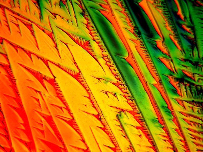
Another blood pressure medication which has the unfortunate side effect of inducing an occasional, but persistent cough. This is Linsopril mixed with Metformin.
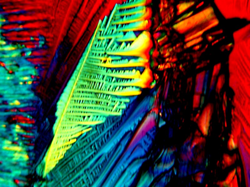
The above is a mixture of Metformin and the very useful stain Alizarin Red S which is an indicator for calcium and some splendid skeletal preparations have been made of small fish by rendering the tissue transparent and then staining with Alizarin Red S.
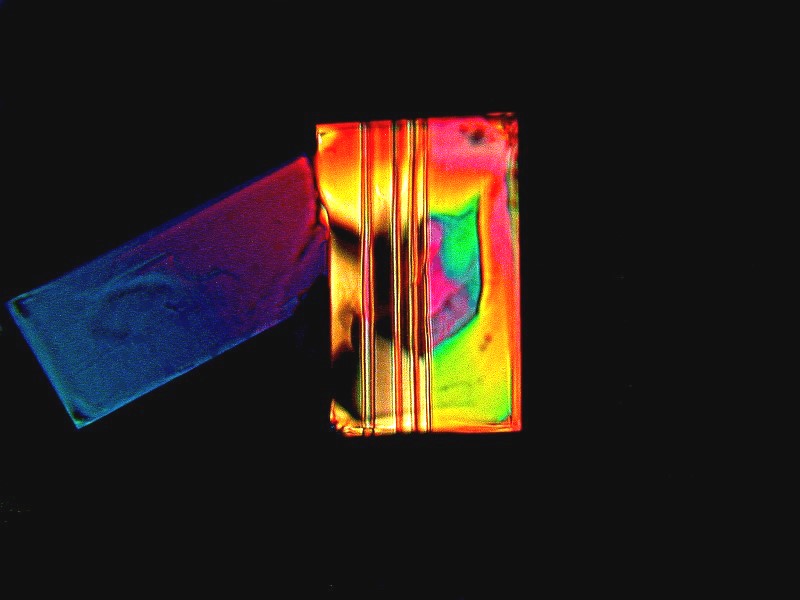
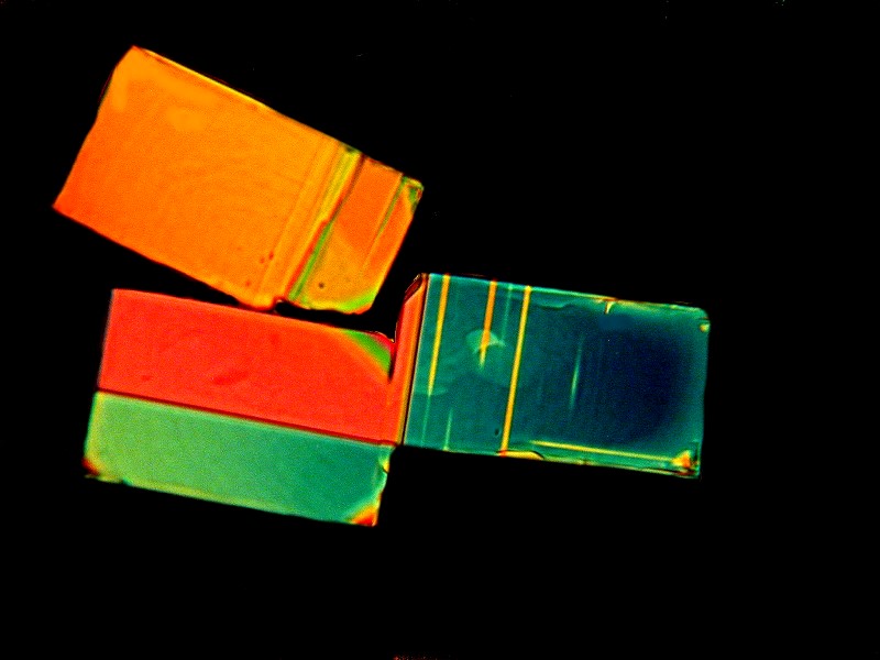
These 2 are a mixture of Metformin with Magnesium chloride which both a delight and a pain to work with. It is a pain because it is hydrophillic and pulls moisture out of the air and the crystals can alter while you are in the process of trying to photograph them. They are delight because the produce wonderful geometric forms, tiny, but magnificent. I did an entire series of images based on Magnesium chloride crystals, in part, because many of them reminded me of the abstract paintings of Mark Rothko in his middle period. Here is a link to my images.
Next comes the glass sponge period. First came the fascination with the Venus Flower Basket (Euplectella aspergillum) about which I have written too much already, so I’ll just refer you to those articles: Euplectella, Euplectella 2, Euplectella 3, Euplectella 4.
Then came a brief adventure (I still hope to do much more) with a glass sponge “with a tail”, Hyalonema. It may sound strange to talk of sponges as “elegant”, “beautiful”, and “stunning”, but just take a look at these examples and you’ll see what I mean.
The spicule types of the hexactinellids (glass sponges) are wonderfully various and I still want to do a brief catalogue of the spicules types of some of these extraordinary creatures.
More recently, I had my chiton period that got interrupted, but I still hope to go back and do some further poking about. I am particularly interested in the 8 dorsal plates and the radulae. What little I did learn, you can find here: Chiton, Chiton 2, Chiton 3 (teeth).
Now, since we are in the prolonged process of moving to a different house (one level–no stairs!), right now until my lab gets set up again I have no immediate access to my microscopes which is, to summarize it in one word–MADDENING! However, once we get settled in to our new residence, I very much look forward to developing some new obsessions and refining a lot of old ones.
All comments to the author Richard Howey are welcomed.
Editor's note: Visit Richard Howey's new website at http://rhowey.googlepages.com/home where he plans to share aspects of his wide interests.
Microscopy UK Front
Page
Micscape
Magazine
Article
Library
© Microscopy UK or their contributors.
Published in the May 2019 edition of Micscape Magazine.
Please report any Web problems or offer general comments to the Micscape Editor .
Micscape is the on-line monthly magazine of the Microscopy UK website at Microscopy-UK .
©
Onview.net Ltd, Microscopy-UK, and all contributors 1995
onwards. All rights reserved.
Main site is at
www.microscopy-uk.org.uk .