 |
ALTERNATIVE MOUNTING MEDIA REVISITED |
|
|
| |
ALTERNATIVE MOUNTING MEDIA REVISITED |
|
|
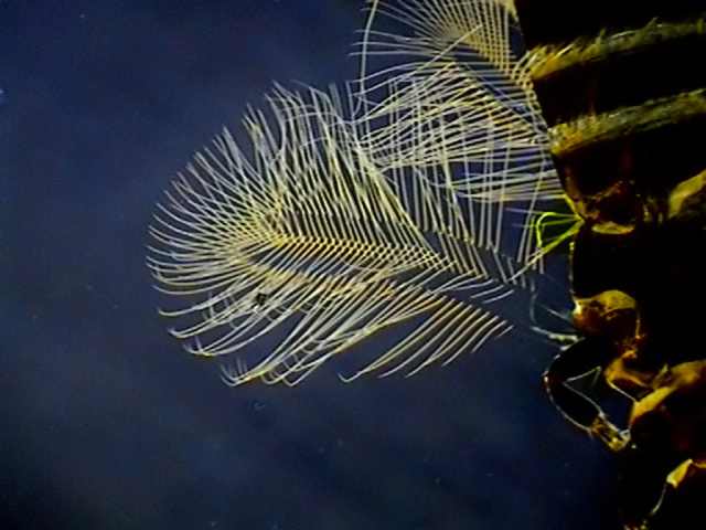
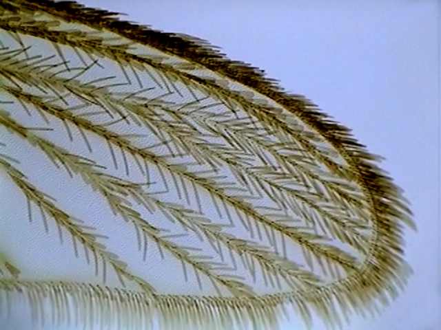
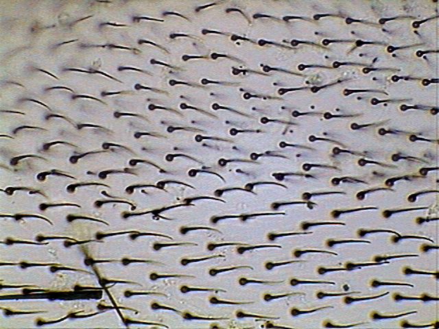
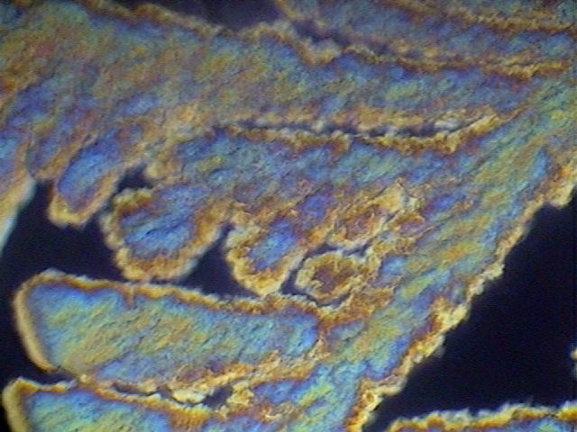
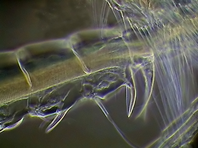
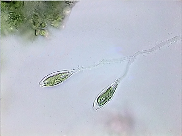
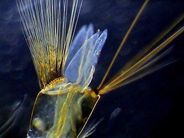
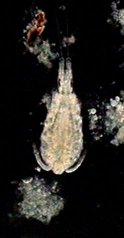
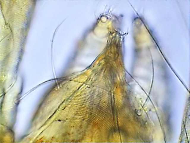
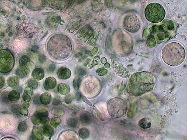
Brachionus
sp.
in a cluster of micro algae. It was fixed with
Lactocupric and mounted in Glycerin
24 months ago. Rheinberg illumination (black center, clear green
periphery). The
illumination emphasizes the mastax trophi and the architecture of the
lorica.
Click on the image to
see it full size.
|
Pictures were taken at 640 X 480 with a Motic DC-3 camera, integrated in my National Optical Microscope, and reduced or cropped for their insertion in the article. Some pictures can be clicked on to see a bigger version. The width of the picture with each objective (on the pictures not cropped of course) is as follows: 4x = 3400, 10x = 1333, 40x = 340 and 100x = 133 microns. Equipment and the filters used were described in a previous article. I wish to
specially recognise the author of the small but splendid Neat
Image Demo
software,
and to Jean
Marie Cavanihac who
brought it to my attention.
INTRODUCTION
Due to the real concern about
the medical
risks associated with the
chemicals used in microscopy laboratories, I presented and
discussed in a
series of Micscape articles (see below) 20 safe mounting
mediums. These
20 formulas provide mounting mediums for all the materials which may be
of interest to the amateur. I present here a critical synopsis of the
most important
results, more
than 20 months after first using the majority of them, and
my selection of the most useful ones. Leaving apart the Damar Gum,
all
the mediums I review do not need any hydrocarbon solvents and therefore
have two main advantages:
1) they are safe, 2) they are cheap. As I stated in my Conclusions
for the series: Leave the Canada Balsam for the professional
taxonomists who
seek legitimately the HYP, (hundred years of permanence) for the
preparations
of their type species, and have many moments of recreation by making
your
collection of preparations look really professional.
www.the
microscopy-uk.org.uk/mag/artdec02/wdmount2.html Part 2. -
Simple drying mediums: www.the
microscopy-uk.org.uk/mag/artjan03/wdmount3.html Part 3. -
formulas: Fructoglycerol and modified
Brun's medium:
www.the microscopy-uk.org.uk/mag/artmar03/wdpart3a.html Part 3b:
PVA-lactic acid and PVA-glycerol
mountants: www.the
microscopy-uk.org.uk/mag/artapr03/wdpart3b.html Part 3c:
The gum arabic mediums www.the
microscopy-uk.org.uk/mag/artmay03/wdpart3c.html Parts 4
and 5. - The glycerin jellies and conclusions: www.the microscopy-uk.org.uk/mag/artaug03/wdpart4.html With
some inevitable repetition, my actual
opinion
on the use of those formulas is presented here..
Indicated when you want to
examine and
preserve for future
examination under the microscope your collections of micro
invertebrates and
micro algae, mixed (or not) with very fine detritus. Fix them using 20%
of Gala
20 or Lactocupric
added to your sample in the field. For a short term
examination you dont need to wash the samples. Try to obtain a very thin
preparation.
Normally it is used for a provisional mounting
planned to last for weeks or some months at the maximum. But because it
allows pictures
to be taken easily, and because the elasticity of the coverslip lets
you move
and turn the individuals observed by applying light pressure with a
dissection
needle, it is one of the most useful techniques that the amateur must
master. Aw mounts
are a very useful for the
rapid screening of a new
sample. As you see, it is only a
refinement, but a
very useful one, of the wet-mount
method. By its nature, even well sealed, the duration of these
preparations is only two or three months. Because of this there are no
examples here. Many
advantages of this technique are
shared by Glycerol, Fructose
or PVA-G (see below) employing in this case
a stepwise technique to
avoid the excess of osmotic pressure that these products can exert on
the micro
invertebrates, all very fragile, and obtaining with a supplementary
effort, a
permanent preparation.
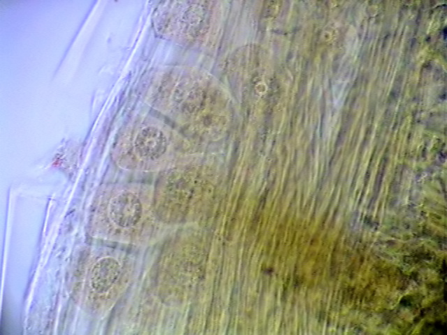
Glycerin is a difficult, but
very useful
permanent mounting medium. It
is also the standard to mount nematodes. It exerts a great osmotic
pressure on
living biological materials or
even on fixed but delicate ones, having as a result the distortion and
collapse
of the individuals. To avoid this it must be introduced very gradually.
The methods to do this were
discussed in the original article. With materials correctly fixed GP
guarantees an almost realistic aspect of the subjects.
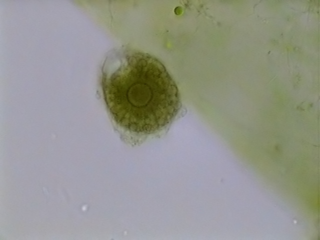
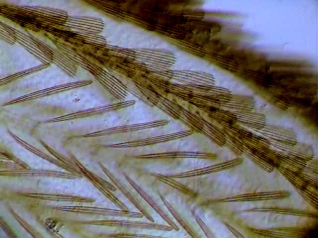
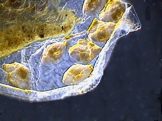
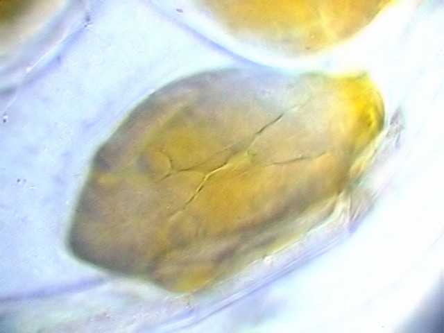
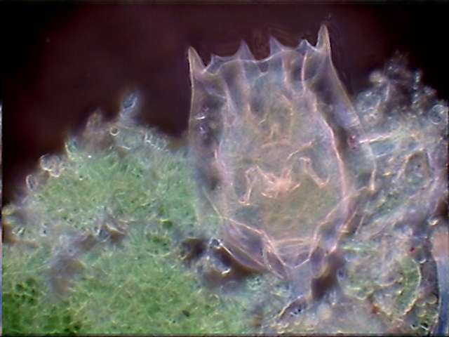
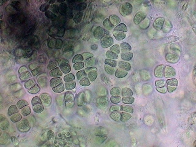
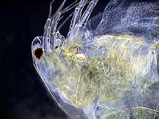
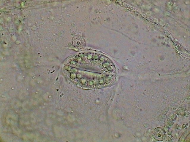
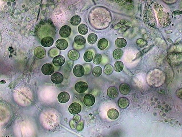
The
pictures show micro-algae
and
some invertebrates mounted in glycerin. The sealing of the
coverslip must be
very carefully done. There
is
always the risk of infiltration. My
preparation
after 24 months has a bubble over almost ¼ surface of the slip.
Clearly 1) my
sealing was not perfect and 2) the materials were not in pure glycerin.
Water has
evaporated. But the remainder of the preparation provided splendid
examples of
the combined effect of lactocupric and glycerin. See later my
discussion of PVA-G.
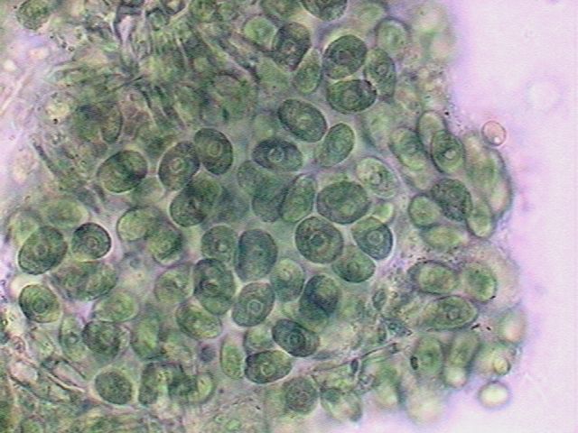
MMF, Mounting mediums with Fructose
Pure, the fructose can be
found as a
powder in the sweeteners section of supermarkets. (If you bought a
bottle (500
g -1
kg), make donations to your colleagues, it will be sufficient for tens
of them
and will last a long time.) This can be
prepared according to the
formula of Larry Legg (see his article in Micscape).
Fructose is suitable for
histological sections
(probably for fungi also) and for many of the invertebrates like small
insects,
entomostracans, or acarina.
Another source,
more convenient and cheap,
is the concentrated corn syrup
(with 76% fructose) which is sold in the
My preparations are thin,
dry, the
coverslips do not move, do not have
any crystallization, and even the
Gentian Violet stain is
preserved. Fructose receives subjects directly
from water, or
glycerin; if you have materials
preserved in alcohol, transfer them gradually to water. Like the
glycerin,
the fructose is capable of collapsing very sensitive materials. A
microtechnique employing
three or four stages can be necessary. It receives and maintains
many dyes, and
preserves very well chlorophyll
in plant samples which were fixed with GALA or Lactocupric. It is one of the easiest and
safest mediums.
I think that Fructose
and PVA-G should be your
standards.
For handicrafts there
are sold some
transparent and syrupy glues made of PVA
(Polyvinyl
Alcohol, not to be confused with Polyvinyl Acetate - also PVA - the
latter
is the basis of students' glues) it can be bought in handicraft
stores,
and in my experience it is not difficult to find. The product is
transparent
and has the consistency of honey and can be diluted with 30% of water.
Some
selected materials (you must test) can be mounted in this adhesive of 70%
commercial
PVA glue, directly from water or from a 1: 3 dilution of glycerol
in
water. The slides
must be sealed because if they are not, the pure adhesive tends to dry
and peels from the glass in a few months. I do not like the refractive
index
of the pure adhesive, the glycerin improves it a lot (see PVA-G).
You do not know the exact
molecular weight
of the prime materials, or
the exact concentration of the adhesives. But you are amateurs, and if
you buy
a tube of 50 ml of adhesive for one dollar, as I do, you would have PVA
for
your life (buy two if you anticipate a long time life.) I encourage the search for this commercial adhesive because the PVA sold for microscopy is very expensive and difficult to obtain and dissolve. Glues are really very cheap and ready to use.
PVA-G. - Polyvinyl Alcohol with
glycerol It receives
and maintains the color
of the
most usual dyes that the
amateur can employ, and also the chlorophyll of plants. And you can
mount
in it practically all subjects, including pollen (with or without
additional
dyes). I
am a fanatic of PVA-G and recommend that this becomes
your
normal
mounting medium.
For
those
difficult materials such as samples mixed with detritus,
which we discussed in the article on PG, a step by step method
can be used
with PVA-G with very good
results. This gives you a preparation
with the benefits (or better) of a glycerol mount, but solid, dry, and
likely to
be filed
easily and examined even with the oil immersion without problems. I use
a
product labeled "ITOYA" Paper Glue.
Water saturated with
borax........12 ml Glycerin...........................................18
ml This
includes a
polyvinyl product which
has 1% of phenol as
an included preservative; it is an
advantage.
PVA-L. - Polyvinyl
alcohol with lactic acid. This, and the now not
recommended GLG, were
proposed as a replacement for the formulation of Hoyer. Hoyer's
is a powerful clearing agent / mounting medium with arabic gum, chloral hydrate
and
phenol, both
forbidden. The lactic acid clears fast and is very penetrating,
and the
formula was promising, but its refractive
index of 1.43, is
disappointing and much lower than that of
glycerin (1.47). The PVA-L is a good clearing medium
which
acts quickly, but if PVA-G is
allowed time, it must be more
powerful, for
the same materials. Of course, the preparations which I have, with PVA-L,
are
dry, firm, and clear. Certain materials like aphids cleared up
excessively,
for example. But some of the slides had an additional problem which I
do not
know
what to ascribe it to. They had small crystals strewn under the
coverslips.
For the moment I must
recommend the use of PVA-G, and to continue
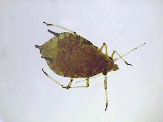
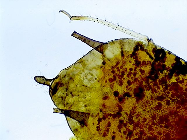
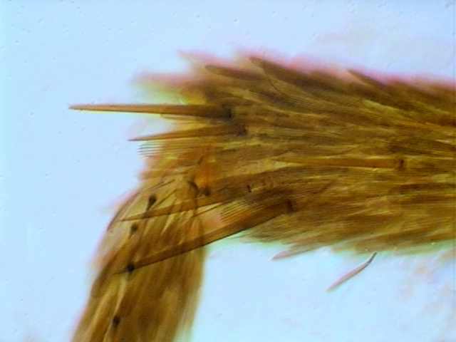
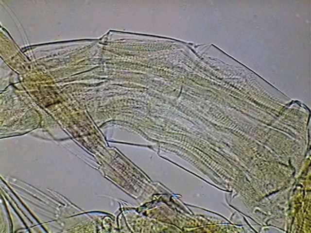
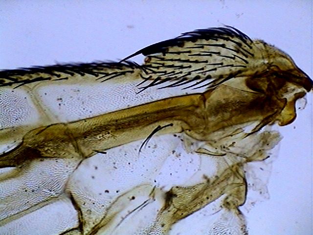
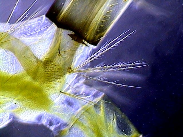
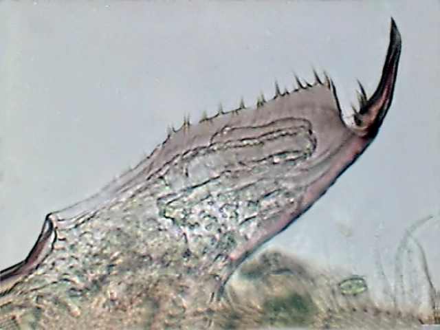
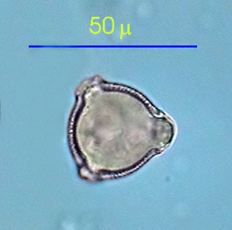
some experiments with PVA-L. Here two successful examples of
mountings with
PVA-L. The
left-hand
picture is from a small
"thrip" (an insect of the order Thysanoptera) captured on a Gerbera jamesoni flower. It
was fixed with
alcohol 70%, washed in water and mounted with PVA-L, 8 months
ago.
The preparation
is perfectly dry, firm and clear, without crystals. Notice the
particular wings of this insect. Obj. X10. Illumination through the
COL-D3. Six
pictures were stitched for this image. The background was blurred
with
Photo
Paint.
The common solvent for all
nail
varnishes is
acetone, butyl acetate or many
other commercial solvents. Generally, to employ it as
a mounting medium, it is wise to dilute it a
little with its solvent to make sure that it runs well under the
coverslip.
I have
mounted leaf epidermis,
hairs,
scales of butterfly, and parts of
insects, algae, protozoa and rotifers. The worst subjects were the
cylindrical
branches of filamentous algae, which were flattened by dehydration with
alcohol.
Being
a natural
resin of vegetable origin, Damar shares with balsam the property
of long
life. It is almost a HYP. Read the
description of
its preparation and techniques for use in the previous article.
It
is naturally a very good choice, but
for the adult amateur. There
is a protozoologist who recommends it as the best medium to mount gregarines
(a form of parasitic protozoa).
II raised
my objections to this much advocated
medium in the previous
article. Nothing has changed.
The Fishers
borax jelly is the most difficult formula,
although it is much
recommended (for pollen especially). Even with double borax and
sufficient
disinfectant, this formula becomes easily soiled with fungi and
bacteria. I do
not use it.
See my comments and
preparation
techniques in the corresponding
article. Apathy was always blamed for the
crystallization of saccharose. In the very thin
preparations of tissues colored with
The formula does not dry
fast, but it
dries. The subjects included are
well preserved. The only problem is the crystals. Try to seal the
margins.
I've
noted
that GAF, which
incorporates fructose (I use Karo, see the
formulas), does not suffer the
crystallization problem. GAF
is my formula to replace the Lillie's
medium,
derived from Apathys
by incorporating fructose in
place of saccharose.
The results are comparable with PVA-G, even for the
sensitive
protozoa. But the drying of the edges is much slower and in wet
climates could
require the assistance of a furnace. But it does dry.
But,
before you
engage in GAF
preparation and its use, you must remember
that the results are very similar and no better than mounting in fructose
or in PVA-G. If you find Karo or fructose in
your
supermarket you do not need GAF. It is the same one if you
obtain PVA.
I am not a diatomist.
But it is clear to me that this synopsis
must include this synthetic resin which has a refractive index of 1.58,
making it useful for the mounting of diatoms. It is sold to repair
cracked front
lamp glasses of cars. Probably the cyanoacrylate resins which must be
UV
treated
(the intense indirect light of the sun is a good source of UV) could
have more
or less the same index. I never have used it but it is frequently
recommended
by
serious amateurs.
AG, alcoholic glycerol GLG, arabic gum, lactic acid
and glycerin FJ, Fructose Jelly They all have a good
performance
and are
useful
for short term work. But they
do not harden at all, they need to be very carefully sealed, and by no
means are
they better than those discussed above. Ive discarded their use. SUMMARY Thus, you have a useful and
required medium for temporary mounts: Aw.
Four
mediums of choice for the average amateur: FMM (home made solution of
fructose, or preferably commercial fructose syrup of the Karo type), PVA-G, NPM, and the Kaisers Jelly. And a medium
for adults: Gum Damar.
If you
can not find fructose or PVA, use the very good but a little more
difficult GAF. Two good
alternative
mediums: PVA-L, for a
similar result to the PVA-G; PG,
difficult
to seal, for certain subjects it is similar to PVA-G, (for the moment
PG must
be considered the standard for the nematodes, in the future we must
compare it
with the PVA-G or the PVA-L). We
also have a special medium for diatomists: Norland 61. So the present review reduces
the list of
the most useful
amateurs' safe mounting mediums from twenty to nine (postponing my opinion
on PVA-L pending additional experiments). References
Marcel Lecomte site http://users.skynet.be/Champignons_passion/main.htm Jean Legrand CONSTRUCTION D'UNE HOTTE DE SECHAGE http://www.microscopies.com/DOSSIERS/Magazine/Articles/JLegrand-Hotte/Etuve.htm
|
||||||||||||||||||||||||||||||||||||||||||||||||||||||||||||||||||||||||||||||||||
Please report any Web problems or offer general comments to the Micscape Editor.
Micscape
is the on-line monthly
magazine of the Microscopy
UK web
site at Microscopy-UK


 There is
no chemical
reason which I can
discover that justifies this
fact and I believe simply that one needs to be very neat when one
washes the coverslips,
the slides, and also sourcing the fixatives or other reagents added to
the
subject.
There is
no chemical
reason which I can
discover that justifies this
fact and I believe simply that one needs to be very neat when one
washes the coverslips,
the slides, and also sourcing the fixatives or other reagents added to
the
subject.