Key: italics: text for experimenters blue:
keywords red: beware!
2. A BRIEF HISTORY
 The
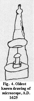
invention of the microscope was not sufficiently documented in its time
to permit a definitive conclusion as regards date and inventor, but the
first illustration of a recognizable microscope dates back to 1625 (Fig.
4). In the 17th century, "compound" microscopes
consisting of a combination of lenses in two groups: objective
(close to the object) and eyepiece (what you
look through) were used by many observers, but the image quality was poor
- comparable to Fig. 1, left.
The
invention of the microscope was not sufficiently documented in its time
to permit a definitive conclusion as regards date and inventor, but the
first illustration of a recognizable microscope dates back to 1625 (Fig.
4). In the 17th century, "compound" microscopes
consisting of a combination of lenses in two groups: objective
(close to the object) and eyepiece (what you
look through) were used by many observers, but the image quality was poor
- comparable to Fig. 1, left.
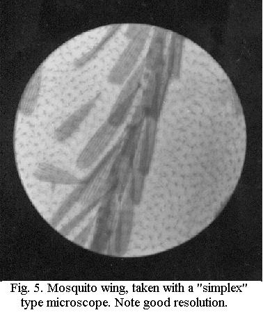
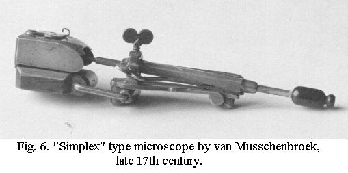
There was an alternative: the "simplex"
microscope consisting of a single lens, like the magnifiers that
are still in use, but of much higher power. This was the type of instrument
used by Anthonie van Leeuwenhoek around 1700 and at the same magnification,
this yielded a much better image than did the compound microscope then.
Fig. 5 shows the image quality obtainable with a "simplex" microscope (or
"high-power magnifier") which I made myself. The disadvantage of this type
of microscope is that it's almost impossible to work with: like Leeuwenhoek's
crude gadget, the beautiful slightly later instrument by van Musschenbroek
(Fig. 6) is hand-held, almost pokes into the user's eye and the observer
must try to look through a tiny (about 2 millimetres) lens. This was the
main reason why the astounding range of van Leeuwenhoek's discoveries
- from striated muscle to bacteria, described in over 560 Letters to the
Royal Society in Great Britain - did not result in the large-scale introduction
of the "simplex" microscope for research.
(A single-lens microscope
has been traditionally called a "simple" microscope, but I have intentionally
changed this to "simplex" microscope to avoid confusion with the
simpler type of modern microscope, e.g. a student's microscope).
The shortcomings in image quality
- especially loss of fine detail due to the presence of colour
fringes - were also known from the telescope. At the end of the
18th century it was discovered that a telescope objective consisting of
a combination of lenses made of different kinds of glass could greatly
reduce the colour fringes, leading to a revolution in optics. Such lenses
were called "achromatic", free of colour.
In the beginning of the 19th century, this principle was also applied to
the objective of the compound microscope. Together with theoretical and
empirical studies by investigators like Lister in Great Britain and Amici
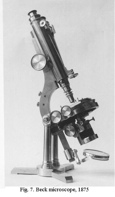
in Italy this resulted in continuous improvement. From about 1850 to 1870,
British instrument makers like Power & Lealand, Ross and Beck led the
field and these Victorian microscopes (Fig. 7), with a tubelength of 10
inches, were truly monumental examples of the instrument makers' craft.
Diatomists wanting to resolve ever finer detail created a genuine demand
for optical improvement and thus formed an important market.
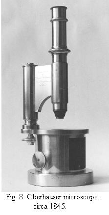
On the European Continent,
a simpler and smaller (tubelength around 160 mm) model (Fig. 8) had become
the standard and by 1870, optical performance of the lenses produced by
European opticians like Hartnack had caught up with the British. The German
instrument maker Carl Zeiss then joined forces with a physicist, Ernst
Abbe, who was the first to create a comprehensive theory
of image-formation for the microscope. Combining the earlier work
with his own studies, Abbe developed a rigorous mathematical treatment
of lens design. Together with the development of new types of glass, this
raised the resolving power of the microscope to the limit of what is physically
possible. Abbe's work was in the public domain, as we say now, so that
all designers could use his data to perfect their own products. Since circa
1890 images have become crisper thanks to coatings
and the field of view has become larger and
sharp almost to the edge by "wide-field" optics,
but the resolving power of the classic LM
has remained the same. For much higher resolving power the electron-microscope
has become available.
Another important 19th-century development
was the mass-production of perfectly clear cover-glasses of uniform thickness
(or rather: thinness, they are only about 1/6th of a mm "thick"). As early
as 1868, one British manufacturer alone sold about 900,000 of them, which
tells a lot about the interest in microscopy at that early time!
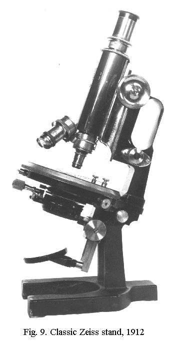
The late 19th-century "Continental"
type of stand (Fig. 9) was the standard for almost half a century. In the
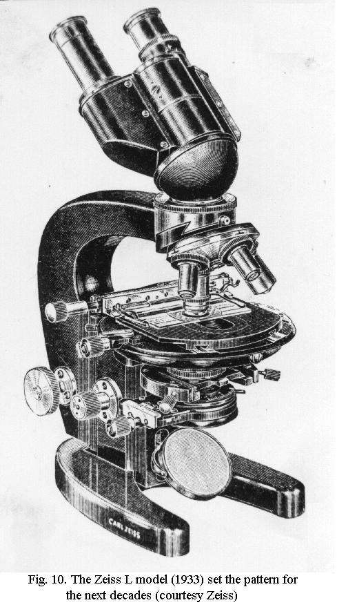
early Thirties Zeiss introduced a revolutionary type of microscope (Fig.
10), which was of the modular type, permitting a choice from a wide range
of different components. Comfortable viewing was assured not by inclining
the stand as in Fig. 9, but by an inclined tube. The model shown has a
binocular
tube for viewing with both eyes - now standard for larger instruments.
Variations on this type of stand have been the standard ever since. Coatings
were introduced just before the Second World War and shortly after it,
two important optical techniques were introduced: phase-contrast
(whose inventor, Zernike, was awarded the Nobel Prize) and interference
contrast. Both make it possible to enhance the visibility of almost
transparent objects like living cells.
«««« »»»»
Hosted by Micscape,
the monthly magazine of Microscopy-UK.
September 2002
© Frithjof A. S. Sterrenburg
© Onview.net Ltd, Microscopy-UK, and all contributors 1995 onwards. All rights
reserved. Main site is at www.microscopy-uk.org.uk with full mirror at www.microscopy-uk.net.