1. INTRODUCTION
The fallacy
For about 350 years, the telescope
and the microscope have enlarged human concepts more than any other scientific
tool. However superficially, the Macrocosm and Microcosm are now part and
parcel of our life - from Star Trek adventures to anthrax bacillus letters.
Yet both the telescope and the microscope have always suffered from the
fallacy that their contribution is thought to consist of "magnification".
The first step towards any understanding of what you're trying to achieve
by using a telescope or microscope is to get rid of this fallacy once and
for all. We'll limit the discussion to the microscope here, but for the
telescope the situation is wholly comparable because both instruments are
subject to the physical laws of optics.
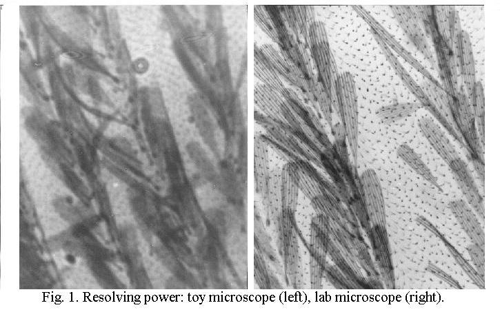
That magnification does not automatically yield additional information should be self-evident now we widely use PC's for images. Simple image-processing software offers the "zoom" tool, but blowing up a picture will merely result in a very coarse-grained image that contains no additional information. The aim of using a microscope is not to magnify an image, but to see finer details in the image. This fundamental difference is clear from Fig. 1, two images of the scales on the wing of a mosquito. Magnification is identical, the left picture was taken with a toy microscope, the right picture with a laboratory-type instrument.
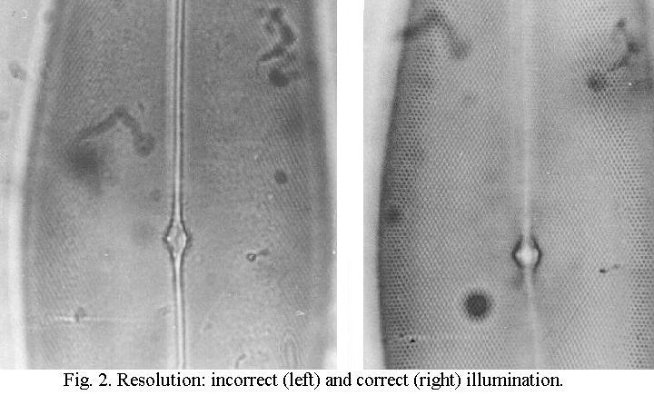
Inferior optical quality is not the only factor that can lead to poor images, as shown in Fig. 2, pictures of the diatom Pleurosigma angulatum. Both were taken with the same set of optics, the difference in quality is exclusively the result of poor and correct microscope handling respectively.
The performance
of the microscope is expressed as its "resolving power", the ability
to separate ("resolve") fine details.
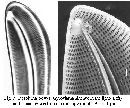
Diatoms in microscopy
Diatoms
are microscopic plants ("algae") consisting of a single cell surrounded
by an exoskeleton consisting of silica. The silica walls have a complex
ultrastructure, which in many species consists of a very regular pattern
of perforations. Fig. 3 shows the image of Gyrosigma sinense in
the light-microscope (LM) and the ultrastructure of this diatom in the
Scanning-Electron Microscope, "SEM". In the LM the image is much less detailed
and is reduced to a series of "dots" arranged in straight lines crossing
at an angle of 90º . The fineness and regularity of this pattern of
"dots" in the LM are the reason why diatoms have been valuable test
objects for assessing the quality of the microscope image since
about 1850. Diatoms can be regarded as a highly representative example
of "a microscopic object", so that we will often refer to them. Diatom
slides are available commercially, see the Internet.
«««« »»»»